
|
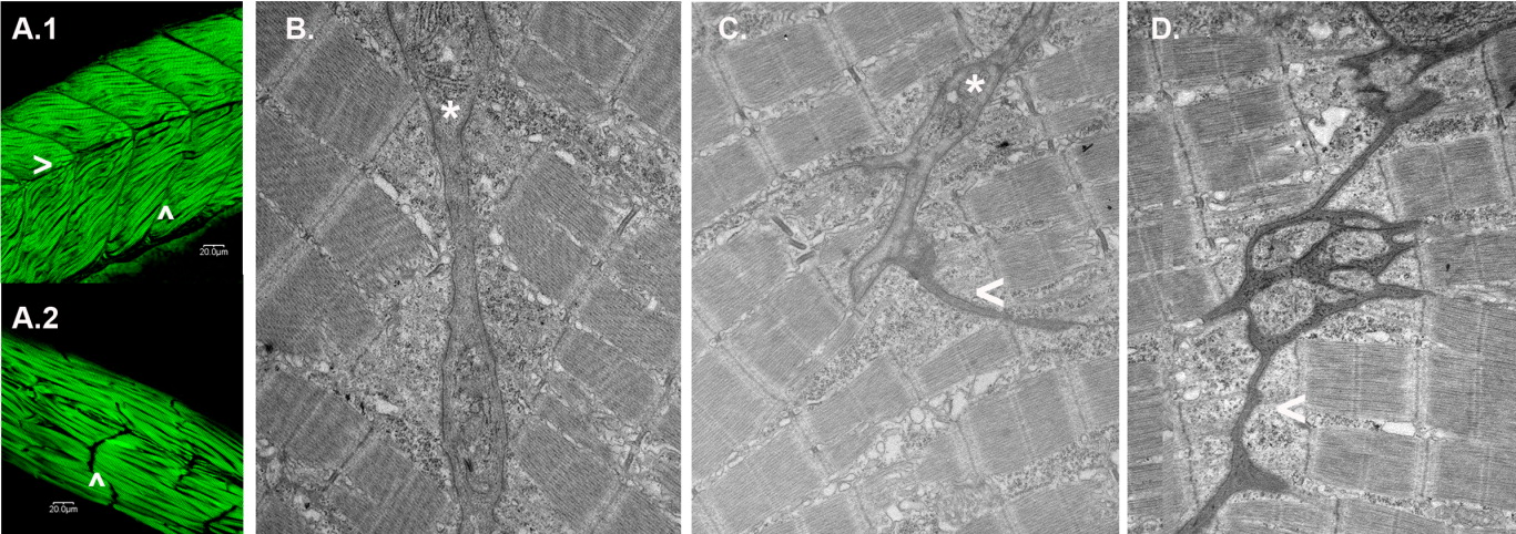
Fig. 6 Obscurin depletion disrupts somite architecture. A: Embryos injected with MO2 (6 ng; A.2) or a control morpholino CMO2 (6 ng; A.1) were fixed and immunostained for α-actinin at 72 hpf. Note that in the morphant embryos, there are no detectable horizontal myoseptae (>) and only rudimentary transverse myoseptae (∧) compared to control embryos. Elongated, disarrayed myofibrils often extend beyond the length of a normal somite. B-D: Electron micrographs of transverse myoseptae from control [6 ng CMO2 (B)] and obscurin morphant [3 ng (C) and 6 ng (D) of MO2] embryos. Morphant embryos displayed rudimentary transverse myoseptae (C:*, C,D: <) at the ends of the skeletal myocytes compared to the well-organized transverse myoseptae of control embryos (B: *).