
|
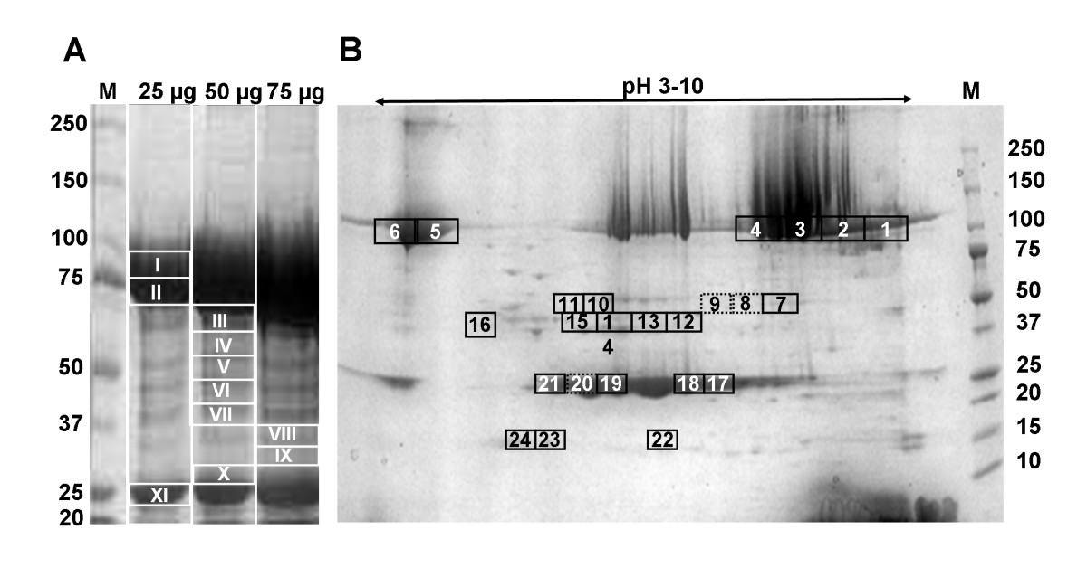
Fig. 4 Electrophoretic pattern of zebrafish fully-grown follicle proteins. A, SDS-polyacrylamide gradient 8-16% slab gel of total yolk proteins with increasing amounts of proteins deposited per lane. Eleven gel slices, numbered I to XI, were cut after staining the gel with See-Band Forte (GeBA). B, Two-dimensional polyacrylamide gel electrophoresis of total yolk proteins. Twenty-four spot areas, numbered 1 to 24, were excised after staining the gel with GeBA. In both procedures, gel pieces were in-gel digested with trypsin and the resulting peptides identified by mass spectrometry (see Table 5). The rectangles from which VTG derivatives were isolated are identified with a solid lined line while the rectangles with no derivatives (numbers 8, 9, and 20) are labelled with a broken line. M, relative molecular weights of the standards x 10-3.