
|
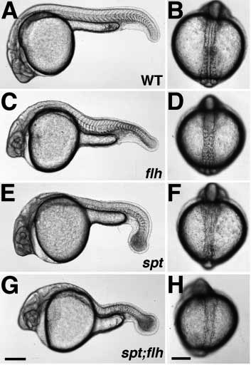
Fig. 1 Phenotypes of live embryos during pharyngula (24 h) (A,C,E,G) and segmentation (13-14 h) (B,D,F,H) stages. In wild-type embryos (A,B), the notochord is flanked on either side by the developing somites, which give rise largely to myotomal muscle. In flh- embryos (C,D), the notochord is missing and the somites (and later, muscle) are fused across the midline. Like spt- embryos (E,F), spt-;flh- embryos (G,H) lack trunk somites, but have tail somites. Although a notochord is present in both spt (E,F) and spt;flh (G,H) mutant embryos, it is incomplete in the double mutants. The embryo shown in H is an example of moderate notochord rescue; almost 90% (30/34) of spt-;flh- embryos have a detectable notochord primordium during early to midsegmentation stages. In about one-third (11/34) of spt-;flh- embryos, notochord rescue is so extensive that double mutant embryos are indistinguishable from spt single mutant embryos, at least at the level of the dissecting microscope. (A,C,E,G and B,D,F,H) Lateral and dorsal views, respectively. Scale bars, 250 um (left panels), 200 um (right panels).