
|
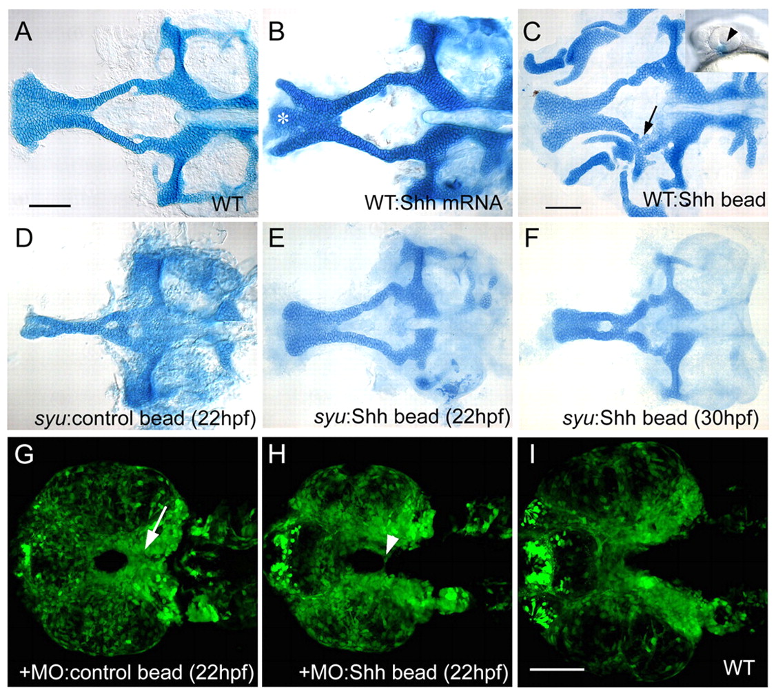
Fig. 7 Exogenous Shh expands ANC cartilage in wild types and rescues syu mutants. Ventral views, anterior to the left. (A-F) Flat-mounted, Alcian-Blue-stained cartilages at 4.5 dpf. (G-I) Confocal images of sox10:egfp expression in dissected embryos at 36 hpf. (A) Wild type. (B) Wild type injected with shh mRNA. Trabeculae expand laterally and often separate from the ethmoid (asterisk). (C) Wild type implanted with a Shh-coated bead on the left side at 20 hpf. The ANC expands near the bead; both trabeculae and ethmoid thicken, and the trabecula on the implanted side fuses to the palatoquadrate (arrow). Inset shows the typical bead position (arrowhead). (D) Control beads implanted at 22 hpf had no effect on ANC formation in syu mutants. (E) Implantation of a Shh-coated bead partially rescues the ANC in syu mutants. (F) Bead implantation at 30-36 hpf also partially rescues the ANC in some cases. (G) Control beads implanted between 22-36 hpf had no effect on the distribution of sox10:egfp+ cells in Hh-deficient embryos. Arrow indicates NC cells at the midline. (H) Implantation of a Shh-coated bead at 22 hpf rescued sox10:egfp expression at the ventral midline (arrowhead). Note that the width of the bead-implanted embryos is identical to that of controls (G,H), and smaller than that of wild type (I). Scale bars: 50 μm; in A for A,B; in C for C-F; in I for G-I.