
|
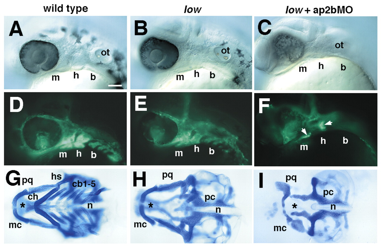
Fig. 5 Cartilage defects correlate with postmigratory NCC defects in low mutants and larvae injected with ap2bMO. Columns consist of photos taken at different stages in the same animal. Lateral views of living fli1-GFP transgenic embryos at 28 hpf (A-F) and cartilage preparations from the same individuals at 4 dpf (G-I). fli1-GFP expression includes both NCC as well as developing endothelial cells. Arrows in F indicate residual NCCs. b, branchial arches; cb, ceratobranchial; ch, ceratohyal; h, hyoid arch; hs, hyosymplectic; m, mandibular arch; mc, Meckels cartilage; n, notochord; pc, parachordals; pq, palatoquadrate. Scale bars: 100 µm.