
|
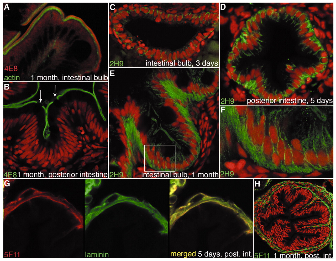
Fig. 3 Monoclonal antibodies that mark brush border, epithelial cell polarity and basement membrane in the zebrafish gut. (A,B) 4E8 antibody stains the brush border, identifying absorptive cells. (A) 4E8 (red) recognises an antigen at the apices of the microvilli, distal to the actin-rich microvillus cores (green). (B) The 4E8 stain (green) is specific to absorptive cells; goblet cells (arrows) are unstained. (C-F) 2H9 (green) stains the lateral membranes and basal regions of gut epithelial cells; the staining is already visible at 3 days, and then becomes progressively stronger and more plentiful basally, especially in the villus epithelium, where cells are basally elongated. (F) Detail of the boxed area in E, showing membrane localization; basal stain may be cytoplasmic, or may reflect labelling of highly convoluted plasma membrane. (G,H) 5F11 labels the basement membrane. (G) 5F11 (red) colocalizes with laminin (green) in the intestinal epithelium at 5 days. (H) At 1 month, 5F11 (green) staining outlines the intestinal epithelium. (B-F,H) TOPRO-3 nuclear stain is shown in red.