
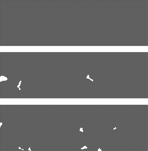
adult female zebrafish - upper image, reference; middle image, 1 nM E2, 3w; lower image, 100 nM E2, 3w Reference ovaries of reproductive female zebrafish (upper image) show a full range of developing and maturing oocytes. After exposure to 1 nM E2 for three weeks (middle), all these maturation stages can still be recognised. However, this exposure dose induced moderate atresia of mature oocytes
 .
.These effects are dose-dependent, an exposure level of 10 nM E2 induced intermediate effects (not shown), and exposure to 100 nM E2 induced severe changes (lower image): only previtellogenic oocytes remain vital, all vitellogenic stages regressed to atretic follicles and subsequent debris
 . This level of E2 exposure apparently inhibits vitellogenesis, presumably through inhibition of gonadotrophin production, and induces maturing oocytes to become atretic. These effects are shown in detail in the next paragraph.
Exposure to these levels of E2 induces increased production of vitellogenin in the liver. This in turn leads to overload of osmo-active vitellogenin in blood vessels, showing as eosinophilic staining plasma
 .
.Female zebrafish exposed to 1 nM E2 are still reproductive, higher levels of exposure induce infertility. In the end, high levels of exposure to E2 are lethal, probably as a result of hemodynamic failure.

adult female zebrafish - 100 nM E2, 3w In ovaries from fish exposed to 100 nM E2 for three weeks, there are many atretic follicles
 with a granulomatous inflammatory reaction
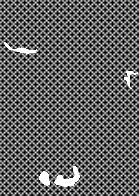 , consisting of macrophages degrading the remains of the non-vital oocytes.
Focally, there are also deposits of lipofuscin pigment
 , consisting of complexes of degraded, peroxidated lipids and proteins.
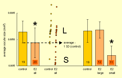
number of included animals are shown in the bars
key findings in the juvenile ovary after estrogen exposure:
- smaller mean oocyte size
 , indicating inhibited development
- speculative presence of two distinctive populations
 , one with normal oocyte sizes (L) and one with smaller oocyte sizes (S); the latter may represent feminized males.
Although delayed ovary development was also concluded after visual characterization (see next image), morphometrical analysis is more precise and enables more detailed conclusions. Oocyte areas were morphometrically determined in 6-weeks old juvenile zebrafish of a control group (control, equal m-f) and a group continuously exposed to 17β-estradiol (E2, all female).
The mean oocyte area in the E2 group is significantly smaller than in the control group
. This suggests that oocyte maturation was inhibited by the E2 exposure.However, the high variance in the exposed group suggests that this is a composite population. An overview of the distribution of cases (middle panel) suggests a normal Gaussian distribution of cases of the control population (most cases within the average +/- 1 SD range
 ), whereas the E2 exposed population contains two clusters of cases: one at the level of the average of the control group, and a second in the lower segment. These populations Large and Small were arbitrarily separated at average - 1 SD (control). Subsequent recalculation yields an E2 population with a similar mean oocyte area as in the control group
 and an E2 population which has significantly smaller mean oocytes than the control and the E2-large groups
 . This speculative distinction between two populations is rational in view of the assumption that the E2 exposed group contains feminized males: E2 exposure might not affect genotypical females, but would transform genotypical males to females with a delayed ovary maturion.

Application of the ovary maturation classification in the evaluation of the effects of effluent of a sewage treatment works (Eindhoven, the Netherlands) in zebrafish.
Zebrafish and subsequent hatched larvae / juveniles were not exposed (control Dutch Standard Water, DSW) or exposed for 6 weeks to 17β-estradiol (E2) or sewage effluent.
Shown is the average of the percentages of the total number of animals in the respective groups (2-5 groups per treatment, 30-50 individuals per group) in each class.
It appeared that exposure of juveniles to either E2 or sewage effluent inhibited ovary maturation, compared to control animals. This conclusion was confirmed by morphometrical analysis of a control and a E2 exposed group (see bar graph above). However, the suggestion of the presence of two subpopulations in the E2 groups, as concluded from the morphometrical analysis, was not apparent from this visual staging, which is therefore considered as less precise.