
adult female zebrafish, control (top), and exposed to 320 µg tamoxifen /L for 3w (bottom) After exposure to the estrogen antagonist tamoxifen, mature and early vitellogenic oocytes in the ovary show various degrees of disintegration
 , compare to normal vitellogenic oocytes
 in the control specimen.
As a result, there are more atretic follicles
 in the exposed animals, and these also have oviducts filled with egg debris
 . These effects were observed in the lowest concentration of exposure (32 µg/L), and increased with dose.
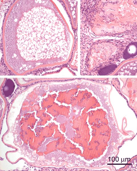
adult female zebrafish exposed to 320 µg tamoxifen /L (3w) These detail images of the ovary after exposure to the estrogen antagonist tamoxifen show how the vitellogenin granules have fused to a intracellular mass
 . Another feature is the accumulation of basophilic granular material
 within the oocytes, possibly disintegrating vitellogenin.
Atretic follicles
 are the end result of this degeneration process.
Associated changes of the granulosa cell layer are shown below.

adult female zebrafish exposed to 320 µg tamoxifen /L (3w) In addition to the changes that occur in oocytes after exposure to tamoxifen (see above), there are changes in the appearance of the granulosa cell layer: granulosa cells are hypertrophied
 , i.e. there is an increased size of the cell and its nucleus, and the latter is lobulated and hypochromatic. This is a focal phenomenon, since normal appearing granulosa cells
 are also present.
Furthermore, invaginations of the oocyte membrane
 are observed, associated with the presence of transformed granulosa cells.
The observed changes occurred from the lowest level of exposure (32 µg/L), and increased in a dose-dependent way.
are observed, associated with the presence of transformed granulosa cells.
The observed changes occurred from the lowest level of exposure (32 µg/L), and increased in a dose-dependent way.This transformed appearance of granulosa cells indicates activation of these cells. This can be understood, since blocking of the estrogen receptors with tamoxifen will induce a negative feedback signal in the pituitary, causing gonadotropin hormone producing cells to increase the release of this GTH. In turn, GTH will stimulate estrogen producing cells, which are the granulosa cells of growing follicles, to increase the production of estrogen.
Note, that a theca layer
 is discernible on both oocytes.
In the lower image, adjacent to the oocyte, an accumulation of transformed granulosa cells, macrophages, and other stroma cells
 is visible as part of an atretic follicle.
See also page on normal granulosa cells.