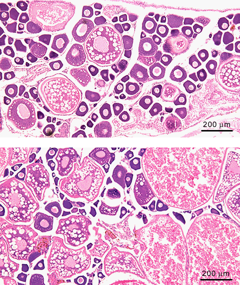
Adult female zebrafish ovary; H&E staining ovary general structure
Two specimen of normal adult zebrafish ovaries, in different stages of the spawning cycle. The upper specimen shows a high ratio of previtellogenic oocytes
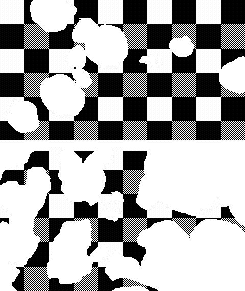
 , suggestive for an early postspawning stage, whereas the lower specimen contains predominantly vitellogenic oocytes
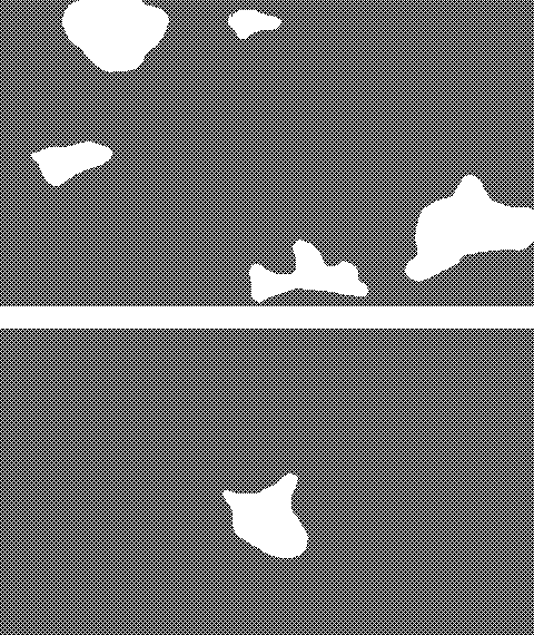
 , indicating an intermediate stage. The postspawning condition of the upper specimen is further indicated by the high ratio of postovulatory follicles
 .
.
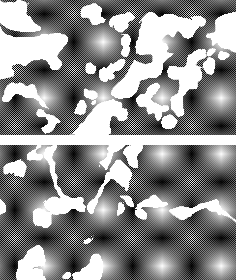
Note, that the upper image shows a lobulated structure, with interlobular spaces
 that communicate with the lateral oviduct
 . Thus, the interlobular spaces give way to ovulated oocytes.

Adult female zebrafish ovary, prespawning stage; H&E staining
In a further (prespawning) stage shown here, mature oocytes have ovulated and are collected in the caudal oviduct
 . This image is suggestive of a prespawning stage.