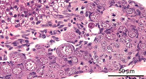
Juvenile female zebrafish, total body length 12 mm; age 4w; H&E staining This image represents an early previtellogenic stage in ovary development, with predominant occurrence of early oocyte stages i.e. oogonium, primary oocytes stage 1, and occasional stage 2.
In this image, the following elements are present:
- clusters of meiotic oogonia
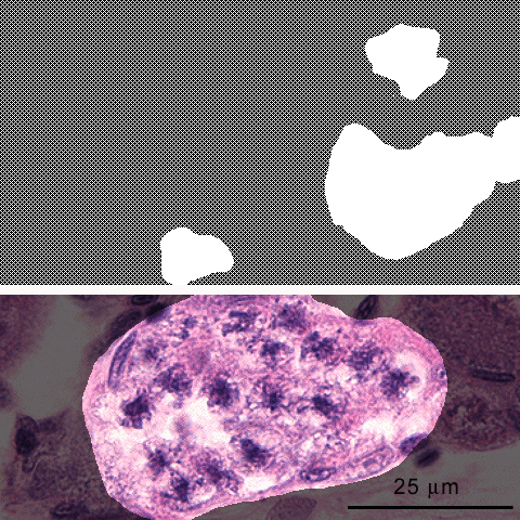 , see below for detail; cell diameters ~10 µm.
 - stage 1 primary oocytes
 : these are small previtellogenic oocytes (cell diameters: 10-20 µm). Accompanying follicle cells
 are present.
 - stage 2 primary oocytes
 : these previtellogenic oocytes have cell diameters ranging of 20-90 µm. Accompanying follicle cells
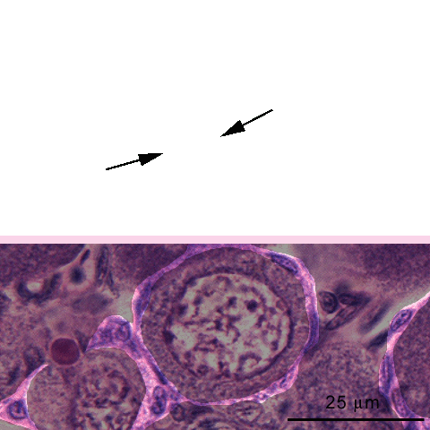 are arranged in a single layer.
 , occasional apoptotic bodies
 are present. The stroma tissue of the mesentery
 between the ovary and the liver
 contains eosinophilic peritoneal cells
 .
.
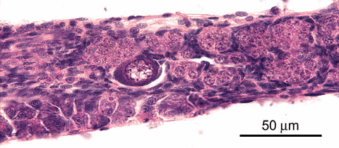
Juvenile female zebrafish, total body length 10 mm; age 5w; H&E staining Ovaries with progressed maturation may still contain only primitive developmental stages, especially in the rostral and caudal poles; this suggests that the growth of these organs emerges from these regions.
In this image, the following elements are present:
- clusters of oogonia
 , (cell diameters: ~10 µm)
 - stage 1 primary oocytes
 , these are the earliest previtellogenic maturation stages of oocytes (cell diameters: 10-20 µm).
 - stroma
- adjacent pancreas tissue

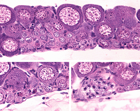
Juvenile female zebrafish, total body length 11 mm; age 4w; H&E staining The images below represent various parts of the same ovary.
The following elements are present:
- oogonia
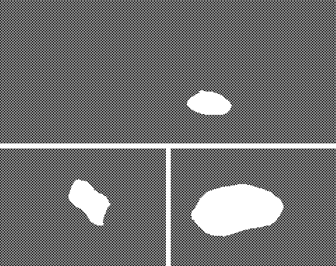 : develop in clusters from germ cells; cell diameters ~10 µm
 - primary oocytes: previtelogenic maturation stages of oocytes; maturation from oogonium is asynchronous. At this age, only stage 1 and stage 2 primary oocytes can be distinguished:
- stage 1 oocytes
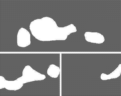 : small, cell diameters range from 10-20 µm; round-oval nucleus with 1-4 nucleoli per section; small rim of cytoplasm; accompanying follicle cells
 are present.
 - stage 2 oocytes
 : increase of cytoplasm volume with corresponding increase in cell diameters to a range of 20-90 µm; the cytoplasm is compact and reacts basophilic. Accompanying follicle cells are arranged in a single layer
 .
.
- stage 1 oocytes
- apoptotic bodies
 .
.
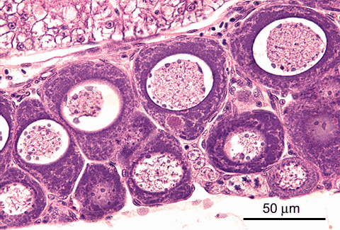
Juvenile female zebrafish, total body length 10 mm; age 5w; H&E staining This image represents an intermediate previtellogenic maturation stage of the ovary. This specimen contains early stages of development up to stage 3 primary oocytes:
- clusters of oogonia
 ;
; - stage 1 oocytes
 (10-20 µm) with accompanying follicle cells
 ;
; - stage 2 oocytes
 (cell diameters: 20-90 µm); cytoplasm is compact and reacts basophilic. Accompanying follicle cells
 are arranged in a single layer;
 - stage 3 oocytes
 (cell diameters: 80-160 µm); cytoplasm reacts basophilic but shows spotted clearing. Accompanying follicle cells
 are arranged in a single layer.
Stroma
 and liver
 are also present.
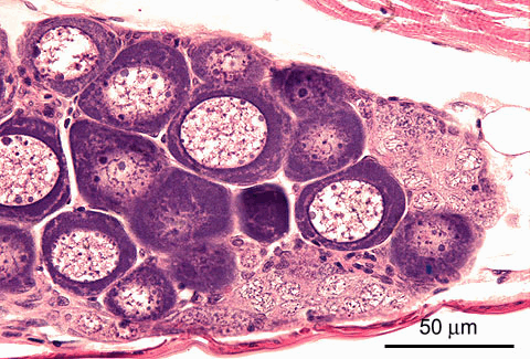
Juvenile female zebrafish, total body length 10 mm; age 5w; H&E staining Generally, further maturing ovaries still have clusters of early oocytes, particularly in the poles of the organ.
Such a region is represented in this image, showing the following elements:
- clusters of oogonia

- stage 1 primary oocytes
 (10-20 µm); accompanying follicle cells
 are present.
 - stage 2 primary oocytes
 (cell diameters: 20-90 µm); cytoplasm is compact and reacts basophilic. Accompanying follicle cells
 are arranged in a single layer.
 - stroma

- trunk wall musculature

- swim bladder wall

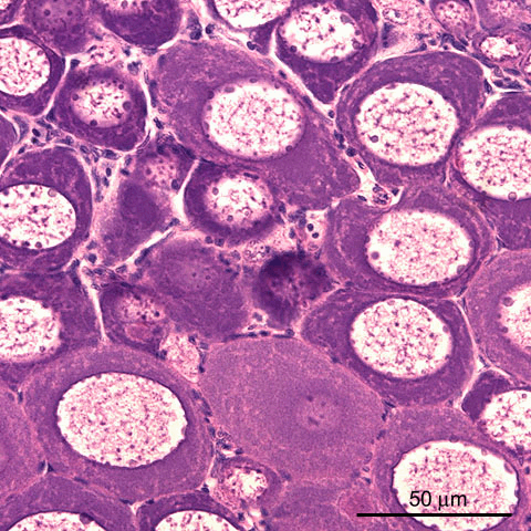
Juvenile female zebrafish, total body length 14 mm; age 6w; H&E staining At advanced previtellogenic maturation stages of the ovary, the section is dominated by stage 2 and stage 3 primary oocytes, with obvious clearing of the cytoplasm. The follicle cells of the stage 3 show more prominent than those of previous stages.
In this image, the following elements are present:
- clusters of oogonia
 ;
; - stage 1 oocytes

(cell diameters: 10-20 µm); - stage 2 oocytes

(cell diameters: 20-90 µm); - stage 3 oocytes
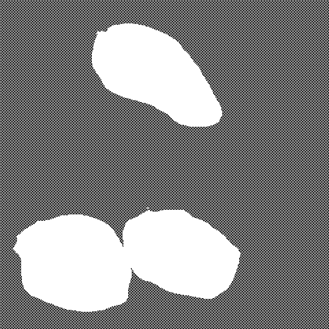
(cell diameters: 80-160 µm); these cells have basophilic cytoplasm basophilic with spotted clearing. Accompanying follicle cells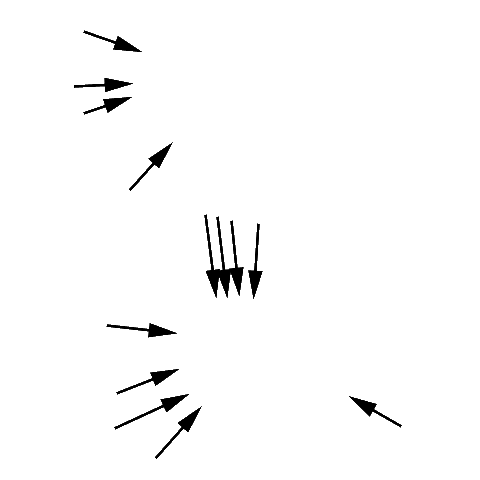 are arranged in a single layer.
 - stroma
