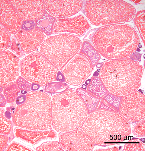
adult female zebrafish, exposed to 10 µg mdhT /L for 3w The non-aromatisable androgen 17α-methyl-dihydrotestosterone (mdhT) has a biphasic effect in the ovary.
After exposure to low or moderate concentrations of mdhT, mature eggs accumulate in the ovary, obviously more than in control animals. Since the eggs are located in the ovary, the androgen apparently has an inhibitory effect on ovulation.
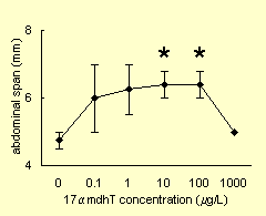
abdominal span measured in adult female zebrafish, exposed to a range of mdhT for 8d This histological observation is supported by measurement of the abdominal span, which increases at the indicated exposure levels, compared to the control animals. At the highest exposure level, the abdominal span is comparable to control. This is explained by the histological image shown in the next section.
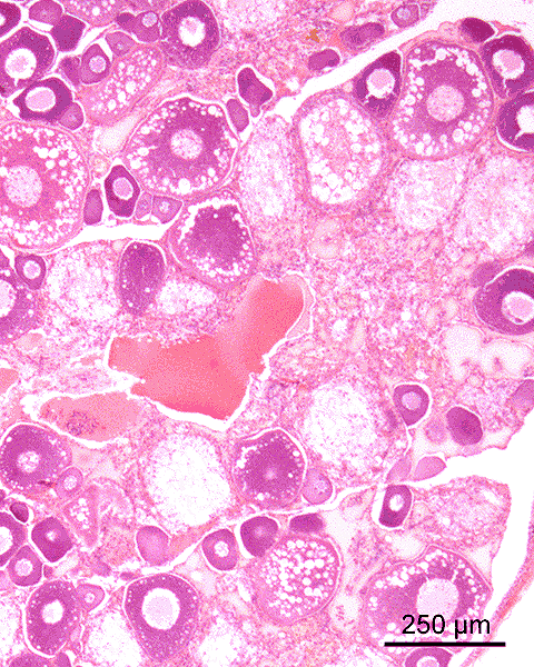
adult female zebrafish, exposed to 1000 µg mdhT /L for 8d After exposure to a high concentrations of mdhT, the ovary contains relatively many non-vital and atretic oocytes
 , compared to control ovaries. As is the case in males, females exposed to such a level of androgen show overexpression of vitellogenin, illustrated by the interstitial edema
 in this image. These effects are similar to those of high levels of estrogen, and can be explained by interaction of the androgen with estrogen receptors.
This image explains why the abdominal span is not increased at a high level of exposure, as is the case at lower levels (see graph above).

After exposure to low or moderate concentrations of the non-aromatisable androgen 17α-methyl-dihydrotestosterone (mdhT) during six weeks after hatching, all juveniles have well-differentiated testes. This effect was only observed after exposure to low or moderate levels of the androgen (0.1 - 1 µg/L), at a higher concentration (10 µg/L) gonads show inhibited development (undifferentiated, or too small to be detected on routine sections).