
- Title
-
Signals from the yolk cell induce mesoderm, neuroectoderm, the trunk organizer, and the notochord in zebrafish
- Authors
- Ober, E.A. and Schulte-Merker, S.
- Source
- Full text @ Dev. Biol.
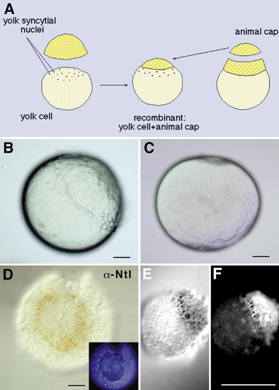
The yolk cell, but not marginal zone tissue, can induce mesoderm in animal cap tissue. (A) Scheme of the experimental procedure. All blastomeres were removed from a high-stage embryo. This yolk cell was then recombined with an animal cap removed from a sibling embryo between high- and sphere-stage. (B) Lateral view of a high-stage embryo, which was incubated in Ca2+-free Ringer’s; animal pole is up. Only the marginal-most blastomeres remain attached to the yolk cell, via tight junctions. (C) Animal view of a completely blastomere-free yolk cell from a high-stage embryo. (D) Animal view of one conjugate. Blastomeres at the periphery of the cap express the pan-mesodermal marker Ntl. (Inset) Animal view of the same conjugate incubated with DAPI showing the external YSL nuclei arranged in a ring-like fashion around the animal cap. The nuclei of the YSL are larger than those of the animal cap blastomeres. (E, F) An animal cap–marginal zone conjugate stained for Ntl protein. The endogenous Ntl expression in the lineage-labeled marginal zone tissue (F) is detected in brown (E), but Ntl expression is not induced in the animal cap explant. Arrowheads point out corresponding nuclei. Scale bars: 100 μm. |

All deep cells are competent to respond to a mesoderm-inducing signal such as activin. (A) Scheme of the experimental procedure. (B) Explants of the central deep cells dissected at sphere stage and cultured until siblings had reached shield stage show no endogenous expression of Ntl. (C) Explants of the central deep cells cultured in activin (8 U/ml) show Ntl expression in all cells. (D) Explants of the marginal blastomeres and animal caps show endogenous Ntl expression only in the marginal explants. (E) Explants of marginal blastomeres and animal caps cultured in activin (8 U/ml) show Ntl expression in all cells. Scale bars: 100 μm. |
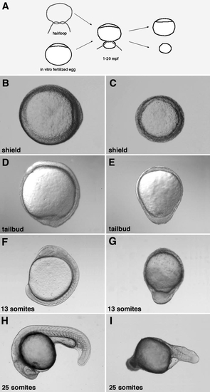
The vegetal part of the yolk cell contains all information necessary to establish dorsoventral polarity and the anterior region of the embryo. (A) The most vegetal part of the yolk cell can be ligated off within the first 20 mpf using a hairloop. (B, C) After 6 h of development morphogenetic movements form a shield at the future dorsal side of an untreated embryo (B), whereas in a ventralized embryo only involution occurs, resulting in a completely radialized embryo. (D, E) Convergence and extension movements lead to the formation of an axis at the dorsal side of the embryo (D), whereas experimental embryos (E) are completely radialized. (F, G) Thirteen somites have formed after 15 h of development in untreated sibling embryos (F), whereas no somites have developed in embryos lacking the vegetal-most part of the yolk cell (G). (H, I) At a stage when the untreated embryo has formed 25 somites, only 11 somites are developed in the experimental case. (B, C) Animal view, dorsal to the right; (D–G) lateral view, dorsal to the right; (H, I) lateral view, dorsal up. |

Gene expression in embryos with lateral (A–D) or vegetal (E–W) yolk removed detected by in situ hybridization and immunohistochemistry. In embryos with removed lateral yolk the expression pattern of zbmp-4 (B) and din (D) is not altered compared to the siblings (A, C). (E, F) zbmp-4 is at 70% epiboly expressed in the ventral epi- and hypoblast and dorsal prechordal plate mesoderm (E), whereas it is ubiquitously expressed in the experimental embryo (F). (G, H) din is expressed at 70% epiboly in the axial mesoderm and dorsal ectoderm (G) and absent in embryos which lack the vegetal pole (H). (I, K) At 30% epiboly nwk is expressed in the blastomeres and the YSL of the future dorsal side (I), but is absent in ventralized embryos (K). (L, M) At 90% epiboly otx-2 is expressed in the anterior-most neuroectoderm (L), but is absent in ventralized embryos (M). (N, O) anf is expressed at 90% epiboly in the anterior-most neuroectoderm (N) and is absent in the experimental cases (O). (P, Q) At tailbud stage, gsc (red, arrowheads) is expressed in the anterior prechordal plate and myoD (blue) in the paraxial mesoderm (P). Transcripts of both genes are abolished in embryos lacking the vegetal pole (Q). At the end of gastrulation sna-1 expression is found in the paraxial mesoderm and the marginal zone (R). In a ventralized embryo paraxial mesoderm expression is absent (S). (T–W) krox-20 (red) is expressed in rhombomeres 3 and 5, gata-1 (blue) marks the blood precursor cells, and Ntl (brown) stains the nuclei of the notochord and the tailbud at the five somite stage in untreated embryos (T, V). In experimental cases (U, W) krox-20 and axial Ntl expression are missing, whereas one can still find Ntl expression in the tailbud. gata-1 is expressed in a radial fashion. (A–H, L, M, and P–U) Lateral view, dorsal to the right (only in R and S is dorsal to the front); (I–K, N, O, V, and W) animal view, anterior to the left. |
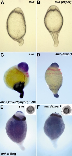
Performing ventralization experiments in swirl embryos rescues neural fates and restores formation of the trunk region. (A) Untreated swr embryo around 13 h of development: the notochord and eight ventrally expanded somites have formed. (B) In an experimental swr embryo a notochord fails to form and the somites are completely radialized. (C) Untreated swr embryo expresses otx-2 (anterior-most staining) most anteriorly, followed by radialized krox-20 (red) in rhombomeres 3 and 5. MyoD is expressed by the radialized somites and brown marks the nuclear localization of Ntl. (D) In experimental swr embryos expression of otx-2, krox-20, and myoD is not changed compared to the untreated swr embryo, whereas axial Ntl expression is absent and only the nuclei of the tailbud still express Ntl. In untreated swr embryos expression of anf (blue) in the anterior-most neuroectoderm and Eng (brown) in the midbrain–hindbrain boundary is radialized, showing a slight reduction dorsally (E). In experimental swr embryos expression of both genes is completely radialized (F); arrowheads mark anf expression. Insets show anterior views of the same embryos; the anf expression domain is pointed out by arrowheads. (A, B) Dorsal view of living embryos, anterior up; (C–F) dorsal slightly to the right, anterior up. |
Reprinted from Developmental Biology, 215(2), Ober, E.A. and Schulte-Merker, S., Signals from the yolk cell induce mesoderm, neuroectoderm, the trunk organizer, and the notochord in zebrafish, 167-181, Copyright (1999) with permission from Elsevier. Full text @ Dev. Biol.