
|
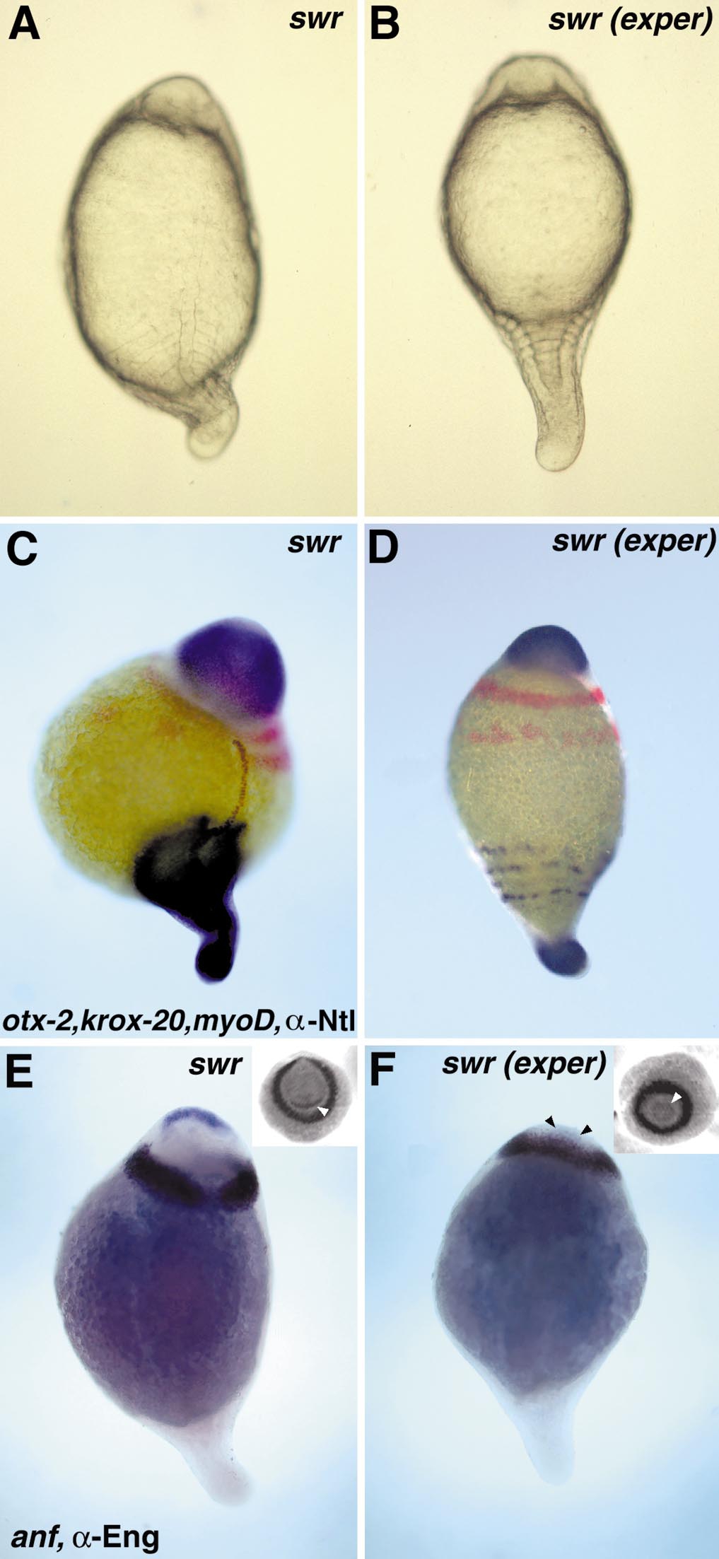
Fig. 5 Performing ventralization experiments in swirl embryos rescues neural fates and restores formation of the trunk region. (A) Untreated swr embryo around 13 h of development: the notochord and eight ventrally expanded somites have formed. (B) In an experimental swr embryo a notochord fails to form and the somites are completely radialized. (C) Untreated swr embryo expresses otx-2 (anterior-most staining) most anteriorly, followed by radialized krox-20 (red) in rhombomeres 3 and 5. MyoD is expressed by the radialized somites and brown marks the nuclear localization of Ntl. (D) In experimental swr embryos expression of otx-2, krox-20, and myoD is not changed compared to the untreated swr embryo, whereas axial Ntl expression is absent and only the nuclei of the tailbud still express Ntl. In untreated swr embryos expression of anf (blue) in the anterior-most neuroectoderm and Eng (brown) in the midbrain–hindbrain boundary is radialized, showing a slight reduction dorsally (E). In experimental swr embryos expression of both genes is completely radialized (F); arrowheads mark anf expression. Insets show anterior views of the same embryos; the anf expression domain is pointed out by arrowheads. (A, B) Dorsal view of living embryos, anterior up; (C–F) dorsal slightly to the right, anterior up.
Reprinted from Developmental Biology, 215(2), Ober, E.A. and Schulte-Merker, S., Signals from the yolk cell induce mesoderm, neuroectoderm, the trunk organizer, and the notochord in zebrafish, 167-181, Copyright (1999) with permission from Elsevier. Full text @ Dev. Biol.