- Title
-
Genetic analysis of fin formation in the zebrafish, Danio rerio
- Authors
- van Eeden, F.J., Granato, M., Schach, U., Brand, M., Furutani-Seiki, M., Haffter, P., Hammerschmidt, M., Heisenberg, C.P., Jiang, Y.J., Kane, D.A., Kelsh, R.N., Mullins, M.C., Odenthal, J., Warga, R.M., and Nüsslein-Volhard, C.
- Source
- Full text @ Development
|
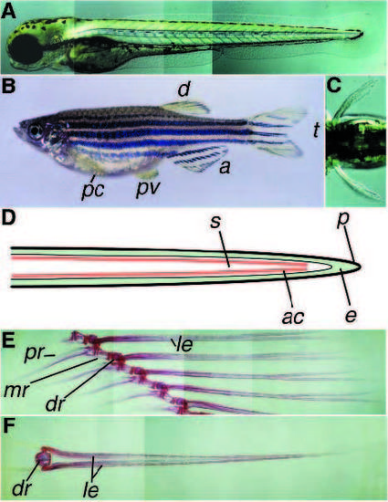
Overview of wild-type fins in zebrafish. (A) Wild-type embryo at 84 hpf, caudal fin. (B) Adult female showing the five different types of fins. Going from anterior to posterior, the pectoral fins (pc) and the pelvic fins (pv), the dorsal fin (d), the anal fin (a) and the tail fin (t). (C) Dorsal view of an approximately 84 hpf wildtype embryo showing the pectoral fins. (D) Schematic drawing of a frontal section through an embryonic fin. Epiderm (e, green) is covered by periderm (p, black), in the subepidermal space (s) between the two epidermal layers actinotrichia (ac, red) are present in a double layer. Actinotrichia are also visible in Fig. 2A. (E) Skeletal staining of a part of the adult anal fin and the supporting skeleton. The endoskeletal radials consist of the proximal (pr), the medial (mr) and a distal (dr) part. The segmented lepidotrichia (le), belonging to the dermal skeleton are connected to the distal radial. (F) Frontal view of one adult anal fin segment showing the bilaterally arranged lepidotrichia connected to the distal radial (dr). |
|
Mutants in which the entire fin fold is affected. (A) Wild-type tail fin lateral view, 48 hpf. Note the thin lines that are running from the base to the tip of the fin, which represent the actinotrichia. Nagel (B), frayed (C), rafels (D), blasen (E) and pinfin (F) mutants, respectively. In nel and pif mutants the bubbly appearance is still visible; later the fin fold will degenerate. (G) Wild-type tail fin, ventral view. (H) frilly fins mutant tail fin, ventral view. The undulations in the fin fold are most pronounced at the tip of the tail. PHENOTYPE:
|
|
Comparison of 3.5-day-old embryos with mutations affecting the pectoral fins. (A,B) Frontal views, (C-I) dorsal views. Wild type (A) and leprechaun (B) mutant embryos showing the increase in pectoral fin size. (C) Wild-type pectoral fins. Both mutations in ikarus (D) and sonic you (E) lead to a variable reduction in the size of the pectoral fins; in D an ika mutant with a clear difference in size between left and right pectoral fin is shown. (F) In dackel mutants pectoral fins are almost invisible at this stage (arrow). (G) Boxer mutants have reduced pectoral fins. The distal parts of the pectoral fins seem to be more affected than the proximal part and show signs of degeneration. This is not observed in ika and syu mutants. (H) Mutant krom embryo showing variably bent pectoral fins. (I) stomp mutant embryo with a degenerating pectoral fin fold. |
|
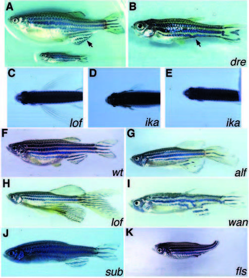
Adult fin phenotype of several of the mutants identified in our screen. (A) Comparison of dre mutant embryo (bottom) with a wildtype sibling (top). (B) Higher magnification of the dre mutant, showing absence of the anal fin (arrow). lof/+; +/+ (C) and two lof/+;ika/ika fish (D,E), exemplifying the variablility of the ika phenotype. (F) Wild-type adult. Both long fin (H) and another long fin (G) heterozygous fish have an increased length of the fins. (I) wanda heterozyogous fish showing strong reduction of the dorsal fin, abnormal pigment pattern and abnormal body shape. (J) Severe example of stein und bein mutant fish. Both pelvic fins are absent and the dorsal fin is severely reduced. In less severe cases the dorsal fin is normal and only one of the two pelvic fins is missing. (K) finless mutant fish; all fins are missing. PHENOTYPE:
|
|
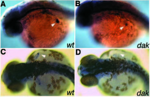
Whole-mount in situ hybridization with sonic hedgehog. Wild type (A) and dackel mutant embryo (B) at 28 hpf. Mutant dak embryos have reduced staining in the pectoral fin buds (arrowheads); no differences were detected in other regions that express this gene (data not shown). Wild type (C) and dak mutant embryo (D) at 32 hpf. In contrast to wild type, no shh expression above background is detectable in the pectoral fins of dak mutants at this age (arrowheads). |
|
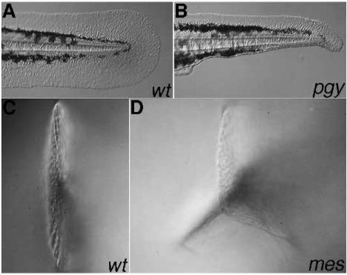
Tail fin phenotype of weakly dorsalized and ventralized mutants. Wild type (A) and piggytail heterozygous embryo (B), side view. The ventral posterior part of the tail fin is missing. Wild type (C) and homozygous mercedes mutant embryo (D), viewed from posterior. On the ventral side of the mercedes tail two fins are visible, giving the tail fin the shape of a star with three arms. PHENOTYPE:
|
|
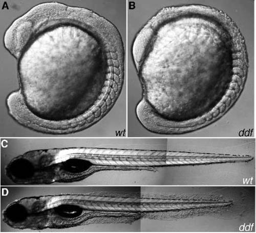
Phenotype of ddf mutant embryos. Wild type (A) and ddf mutant (B) at the 15-somite stage. Rounded-up cells are predominantly present on the head and the prospective hindbrain. The head is apposed more closely to the yolk compared to wild type. (C,D) 5-day-old wild type and ddf mutant, respectively. Rounded-up cells are now easily visible in the fins. The angle between head and trunk appears normal at this stage. PHENOTYPE:
|

ZFIN is incorporating published figure images and captions as part of an ongoing project. Figures from some publications have not yet been curated, or are not available for display because of copyright restrictions. PHENOTYPE:
|

ZFIN is incorporating published figure images and captions as part of an ongoing project. Figures from some publications have not yet been curated, or are not available for display because of copyright restrictions. PHENOTYPE:
|

Unillustrated author statements PHENOTYPE:
|

