- Title
-
Do variants in the CDH23 gene cause non-syndromic retinitis pigmentosa? Dual validation using whole exome sequencing and a zebrafish model
- Authors
- Fan, X., Yuan, S., Wen, J., Wang, S., Rong, W., Feng, D.
- Source
- Full text @ BMJ Open Ophthalmol
|
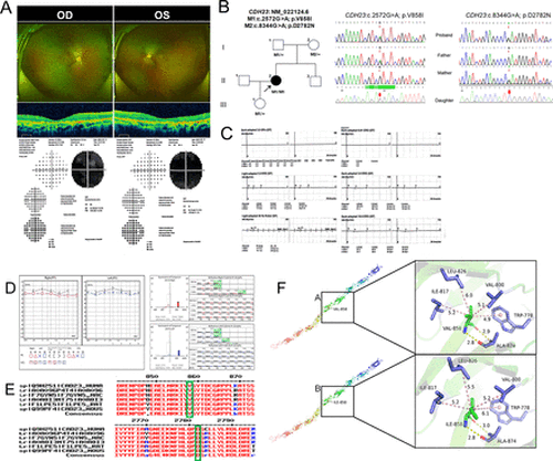
Mutation sequence analysis and clinical examination of the family. (A) Fundus photographs, OCT scans and perimetry results of both eyes of the patient. (B) Pedigree of the family: The filled black symbol represents the affected member, and the arrow denotes the proband. (C) ERG: Significant impairment of cone and rod cell function in both eyes. (D) Pure tone audiometry results of the patient. (E) The homology of amino acid sequences between human CDH23 and other species; the amino acid at positions 858 and 2783 are highly conserved among species, and the mutated residues 858 and 2783 are boxed and indicated. (F) The change in the interaction distance between wild type and mutant proteins further indicates spatial changes. ERG, electroretinography; OCT, optical coherence tomography. |
|
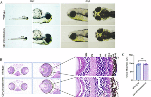
(A) The ocular appearance of wild type and CDH23-knockdown zebrafish at 3 dpf and 6 dpf; (B) H&E-stained retinal sections of wild type and CDH23-knockdown zebrafish at 5 dpf; scale bars: 20 mm; (C) Effects of CDH23 knockdown on retinal thickness in zebrafish. The bar graph presents retinal thickness (μm) for wild type and CDH23-knockdown groups. Data are expressed as mean±SEM (n=5 per group); ns indicates no significant difference between groups (p>0.05, independent-samples t-test) (Representative images shown). GCL, ganglion cell layer; INL, inner nuclear layer; IPL, inner plexiform layer; IS, inner segment; ONL, outer nuclear layer; OPL, outer plexiform layer; OS, outer segment; RPE, retinal pigment epithelium. |
|
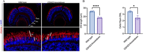
(A) Representative immunofluorescence images of 5-day-old wild type and CDH23-knockdown zebrafish retina. Sections were stained with anti-Alcama (red) and DAPI (blue). Scale bars: 10 µm; (B) Effects of CDH23 knockdown on OS length and ONL cell density in zebrafish retinas. Bar graphs present mean OS length (μm) and ONL cell counts (cells/10 µm ONL) for wild type and CDH23-knockdown groups. Sample sizes: n=5 for OS length and n=3 for ONL cell density. Data are expressed as mean±SEM; *p<0.05, ****p<0.001 (independent-samples t-test) (Representative images shown). DAPI, 4′,6-Diamidino-2-Phenylindole; GCL, ganglion cell layer; INL, inner nuclear layer; IS, inner segment; ONL, outer nuclear layer; OS, outer segment; RPE, retinal pigment epithelium. |
|
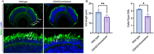
(A) Representative immunofluorescence images of 5-day-old wild type and CDH23-knockdown zebrafish retina. Sections were stained with MPDZ (green) and DAPI (blue). Scale bar: 10 µm. (B) Effects of CDH23 knockdown on OS length and ONL cell density in zebrafish retinas. Bar graphs present mean OS length (μm) and ONL cell counts (cells/10 µm ONL) for wild type and CDH23-knockdown groups. Sample sizes: n=5 for OS length and n=3 for ONL cell density. Data are expressed as mean±SEM; *p<0.05, **p<0.01 (independent-samples t-test) (Representative images shown). DAPI, 4′,6-Diamidino-2-Phenylindole; GCL, ganglion cell layer; INL, inner nuclear layer; IS, inner segment; ONL, outer nuclear layer; OS, outer segment; RPE, retinal pigment epithelium. |
|
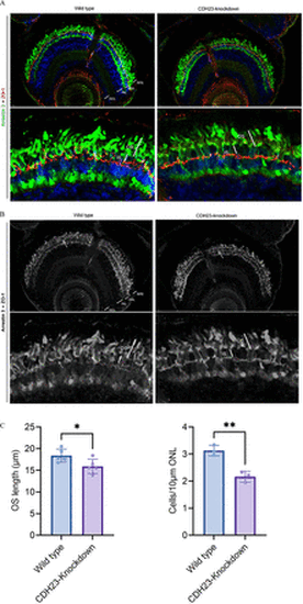
(A) Compared with wild zebrafish, CDH23-knockdown zebrafish photoreceptors are more sparse, but photoreceptors are more closely connected to each other. Up: Scale bars: 10 mm. Down: Scale bars: 2 mm. (B) Grayscale version of panel A; arrows indicate the connecting structures between the photoreceptors. (C) Effects of CDH23 knockdown on OS length and ONL cell density in zebrafish retinas. Bar graphs present mean OS length (μm) and ONL cell counts (cells/10 µm ONL) for wild type and CDH23- knockdown groups. Sample sizes: n=5 for OS length and n=3 for ONL cell density. Data are expressed as mean±SEM; *p<0.05, **p<0.01 (independent-samples t-test) (Representative images shown). GCL, ganglion cell layer; INL, inner nuclear layer; IS, inner segment; ONL, outer nuclear layer; OS, outer segment; RPE, retinal pigment epithelium. |

