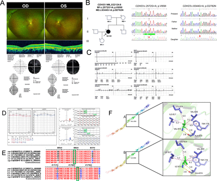
Fig. 1 Mutation sequence analysis and clinical examination of the family. (A) Fundus photographs, OCT scans and perimetry results of both eyes of the patient. (B) Pedigree of the family: The filled black symbol represents the affected member, and the arrow denotes the proband. (C) ERG: Significant impairment of cone and rod cell function in both eyes. (D) Pure tone audiometry results of the patient. (E) The homology of amino acid sequences between human CDH23 and other species; the amino acid at positions 858 and 2783 are highly conserved among species, and the mutated residues 858 and 2783 are boxed and indicated. (F) The change in the interaction distance between wild type and mutant proteins further indicates spatial changes. ERG, electroretinography; OCT, optical coherence tomography.
Image
Figure Caption
Acknowledgments
This image is the copyrighted work of the attributed author or publisher, and
ZFIN has permission only to display this image to its users.
Additional permissions should be obtained from the applicable author or publisher of the image.
Full text @ BMJ Open Ophthalmol

