FIGURE SUMMARY
Title

Homozygosity for a hypomorphic mutation in frizzled class receptor 5 causes syndromic ocular coloboma with microcornea in humans

Authors
Cortés-González, V., Rodriguez-Morales, M., Ataliotis, P., Mayer, C., Plaisancié, J., Chassaing, N., Lee, H., Rozet, J.M., Cavodeassi, F., Fares Taie, L.
Source
Full text @ Hum. Genet.

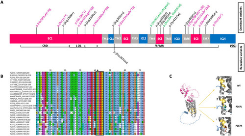
A novel recessive mutation in FZD5. (A) Likely pathogenic variants identified in FZD5. Truncating, missense, and in-frame variants are highlighted in violet, black, and green respectively. The upper section illustrates the position of the dominant (monoallelic) mutations identified in previous reports, while the lower section depicts the novel recessive (biallelic) mutation described in this study. FZD5 contains several important domains: a signal sequence at the N-terminus from amino acids (aa) 1–30; a conserved extracellular cysteine-rich Wnt-binding domain (CRD) from aa 31–150; a linker (L) from aa 151–193; an ordered loop (OL) from aa 194–227; and a Frizzled/Smoothened family membrane region (FSFMR) that includes seven transmembrane Frizzled domains (TM1: 228–260, TM2: 265–297, TM3: 314–345, TM4: 349–379, TM5: 396–428, TM6: 441–479, TM7: 499–522). A PDZ motif is located near the C-terminal at aa 583–585. (EC) extracellular domains and (C) cytoplasmic regions. (B) A multiple alignment was performed for 18 FZD5 orthologs from selected model species, along with all human Frizzled sequences. The YPERPI motif is highlighted (black rectangle); the p.Pro267Leu variant is indicated with an asterisk. Each sequence is identified by its UniProt accession number and the positions of its first and last amino acids in the selection. Amino acids color-coding follows the Clustal scheme. According to the alignment numbering, TM1 and TM2 comprise residues 3–34 and 42–72, respectively. Names of species are the following: DANRE, Danio rerio (Zebrafish); LOXAF, Loxodonta africana (African elephant); MONDO, Monodelphis domestica (Gray short-tailed opossum); PHACI, Phascolarctos cinereus (Koala); SARHA, Sarcophilus harrisii (Tasmanian devil); 9PSIT, Amazona collaria (yellow-billed parrot); NAJNA Naja naja (Indian cobra); ANOCA, Anolis carolinensis (Green anole, American chameleon); LATCH, Latimeria chalumnae (Coelacanth); XENLA, Xenopus laevis (African clawed frog); OCTVU, Octopus vulgaris (common octopus); STRPU, Strongylocentrotus purpuratus (Purple sea urchin); DROME, Drosophila melanogaster (Fruit fly); CAEEL, Caenorhabditis elegans. (C) (Left panel) Alphafold2 model of full-length human FZD5. The color code matches Fig. 1A. The YPERPI motif is highlighted and colored in yellow-orange. (Right panel) Zooming in on position 267 for the wild type (top), the P267L variant (middle), and the P267R variant (bottom). The two prolines of the YPERPI motif are depicted in yellow-orange sticks (P267 and P270), the leucine in deep teal sticks, and the arginine in chocolate sticks. Surrounding residues at position 267, including T339 from helix TM2, N350, and I353 from helix TM3, are shown in grey sticks

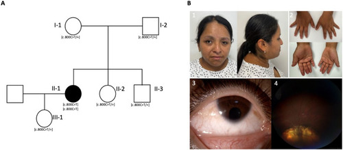
Clinical features associated with the c.800 C > T FZD5 variant. (A)The family pedigree and segregation analysis reveal homozygosity for the NM_003468.4; c.800 C > T variant in the affected individual (II-1) and heterozygosity for the c.800 C > T variant in her parents (I-1 and I-2), siblings (II-2 and II-3), and daughter (III-1). (B) Photograph of the proband (1–4). (1) Demonstrated brachycephaly, facial asymmetry with freckles, strabismus, telecanthus, and epicanthus. (2) Hands exhibiting brachydactyly and slender fingers. (3) anterior segment of the right eye with iris coloboma and microcornea. (4) fundoscopy of the right eye demonstrated retinal and optic nerve coloboma

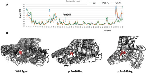
Fluctuation plot and molecular dynamics simulations (A) Fluctuation plot showing the residue fluctuation profile (RMSF) recorded throughout the simulation after global superposition. (B) Molecular dynamics simulations of the wild-type human FZD5, along with the Pro267Leu and Pro267Arg variants, are shown. Ten representative models from the simulation are depicted in cartoon form, with proline, leucine, and arginine illustrated as red sticks

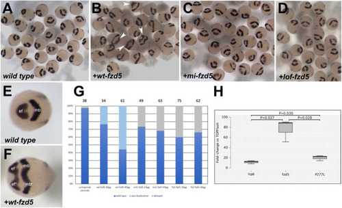
A missense variant in zebrafish Fzd5 mimicking the Pro267Leu variant does not induce a secondary neural axis. (A-F) Expression of rx3 and pax2.1 (dark purple) in 10hpf wild type embryos (A, E) and embryos injected with a wt-fzd5 (B, F), mi-fzd5 (C) or lof-fzd5 (D) mRNA. Arrowheads in (B) highlight some of the embryos displaying double axes. (E-F) Partial or complete double axes are apparent by the duplication of the rx3-positive eye field domain (ef) and the medio-lateral expansion of the pax2.1-positive midbrain domain (mb). (G) Quantification of the effect of injections on body axis establishment. Total number of embryos analysed is displayed on top of the corresponding bars. (H) Fold change in luciferase activity of transfections of lrp6, lrp6 + wt-fzd5 and lrp6 + P277L-fzd5 normalised to activity of TOPFlash alone. Data pooled from three experiments with four replicas each

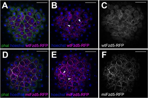
Subcellular localization of zebrafish Fzd5 is not affected by the Pro267Leu mutation. (A-C) Subcellular localization of wt-Fzd5-RFP (magenta, A-B; grey, C) and mi-Fzd5-RFP (magenta, D-E; grey F) in 4hpf embryos injected with the corresponding mRNA. Note the membrane localization of both wt-Fzd5-RFP and mi-Fzd5-RFP (arrowheads in B, E). Embryos were co-labelled with phalloidin-488 to reveal cell outlines (green) and Hoechst to reveal cell nuclei (blue). Scale: 50 μm

Acknowledgments
This image is the copyrighted work of the attributed author or publisher, and ZFIN has permission only to display this image to its users. Additional permissions should be obtained from the applicable author or publisher of the image. Full text @ Hum. Genet.