FIGURE SUMMARY
- Title
-
Detection of acetylcholinesterase and butyrylcholinesterase in vitro and in vivo using a new fluorescent probe
- Authors
- Tang, X., Zhang, Y., Wang, Q., Li, Z., Zhang, C.
- Source
- Full text @ Chem. Commun. (Camb.)
|
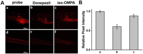
(A) Confocal fluorescence images in zebrafish (head and tail): (a) and (d) zebrafish were only treated with probe OHPD (20 μM) for 4 h; (b) and (e) zebrafish were preincubated with 50 μM donepezil, and then treated with probe OHPD (20 μM) for 4 h.; (c) and (f) zebrafish were preincubated with 50 μM iso-OMPA, and then treated with probe OHPD (20 μM) for 4 h. Scale bar = 100 μm. (B) Relative pixel intensity measurements obtained from the images of Fig. 4A. |
Acknowledgments
This image is the copyrighted work of the attributed author or publisher, and
ZFIN has permission only to display this image to its users.
Additional permissions should be obtained from the applicable author or publisher of the image.
Full text @ Chem. Commun. (Camb.)

