
- Title
-
Protocol for the analysis of hematopoietic lineages in the whole kidney marrow of adult zebrafish
- Authors
- Mahony, C.B., Monteiro, R.
- Source
- Full text @ STAR Protoc
|
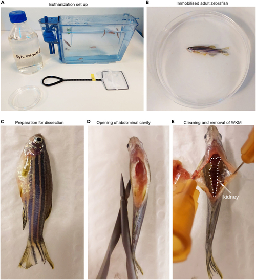
Schedule 1 killing of zebrafish and WKM dissection (A) Euthanization set up and tank of zebrafish. Zebrafish icon by DBCLS (B and C) Immobilized fish. (D) Opening of abdominal cavity. (E) Identification and removal of WKM. |
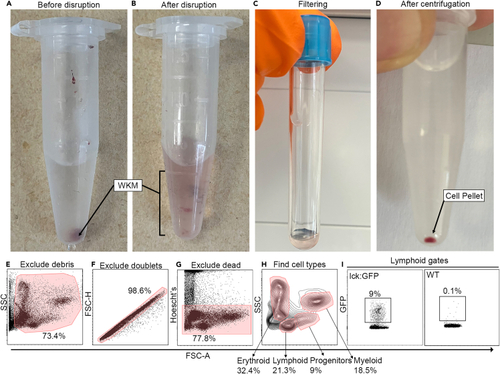
Preparing single cell suspension of WKM for flow cytometry analysis (A and B) Before and after dissociation of WKM. (C) Filtering of WKM. (D) Centrifuging and identification of cell pellet. (E‒I) Gating strategy for analysis. |
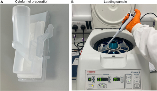
Cytospining cells (A) loading slide into cytofunnel. (B) Loading sample into cytofunnel in cytospin centrifuge. |
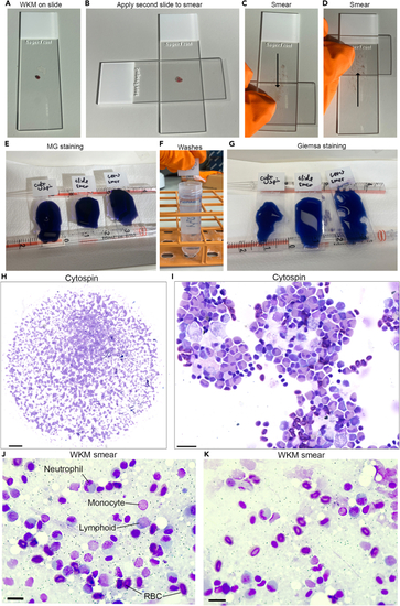
WKM smears and staining cells (A‒D) Smearing WKM on slide. (E‒G) MG staining. (H and I) Cytospun cells. (J and K) WKM smears. Scale bars: H- 500 μm, I- 20 μm, J and K- 10 μm. |