
Image
|
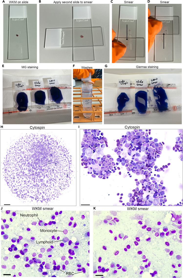
Figure Caption
Figure 4
WKM smears and staining cells
(A‒D) Smearing WKM on slide.
(E‒G) MG staining.
(H and I) Cytospun cells.
(J and K) WKM smears. Scale bars: H- 500 μm, I- 20 μm, J and K- 10 μm.
Acknowledgments
This image is the copyrighted work of the attributed author or publisher, and
ZFIN has permission only to display this image to its users.
Additional permissions should be obtained from the applicable author or publisher of the image.
Full text @ STAR Protoc