FIGURE SUMMARY
Title

Assessment of an Anticancer Effect of the Simultaneous Administration of MM-129 and Indoximod in the Colorectal Cancer Model

Authors
Kwiatkowska, I., Hermanowicz, J.M., Czarnomysy, R., Surażyński, A., Kowalczuk, K., Kałafut, J., Przybyszewska-Podstawka, A., Bielawski, K., Rivero-Müller, A., Mojzych, M., Pawlak, D.
Source
Full text @ Cancers

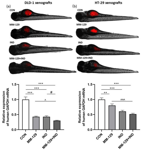
Impact of MM-129 (10 µM), IND (200 µM) or a combination of these agents (MM-129 + IND) on tumor development in DLD-1 (a), and HT-29 (b) zebrafish xenografts, and the expression of human GAPDH mRNA was determined at 120 hpf. Data were presented as mean ± standard deviation (SD) and analyzed using one-way analysis of variance (ANOVA). n = 20 each group; ** p < 0.01, *** p < 0.001 vs. CON; ^ p < 0.05, ^^^ p < 0.001 vs. MM-129; # p < 0.05 vs. IND.

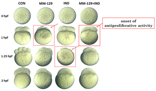
Effect of MM-129 (10 µM), IND (200 µM), and compound combination (MM-129 + IND) on cell division in the zebrafish embryo. Zebrafish eggs after 0, 1, 1.25, and 2 h of exposure to tested compounds; n = 20, hpf: hours post-fertilization.

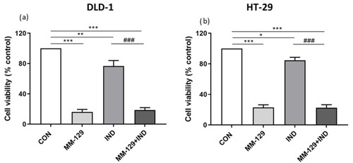
Viability of DLD-1 (a) and HT-29 (b) colon cancer cells treated for 24 h with MM-129 (10 µM), IND (200 µM), and compound combination (MM-129 + IND). Results are presented as mean values ± standard deviation (SD) from three independent experiments performed in duplicate. * p < 0.05, ** p < 0.01, *** p < 0.001 vs. CON; ### p < 0.001 vs. IND.

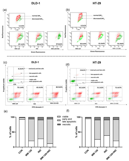
Representative dot-plots illustrating the loss of mitochondrial membrane potential, ΔΨm. (a,b) Flow cytometry dot-plots for the annexin V-FITC-propidium iodide assay (c,d) and quantitative chart illustrating the distribution of live, early, and late apoptotic and necrotic cells (e,f) of DLD-1 (a,c,e) and HT-29 (b,d,f) after 24 h exposition to MM-129 (10 µM), IND (200 µM), and MM-129 + IND. Values were obtained from three independent experiments.

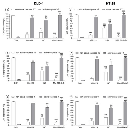
Flow cytometric analysis of caspase-3/7, caspase-10, and caspase-8 activation in DLD-1 (ac) and HT-29 (df) colon cancer cells exposed to MM-129 (10 µM), IND (200 µM), and MM-129 + IND for 24 h. Values from three independent experiments performed in duplicate were presented as mean ± standard deviation (SD) and analyzed using one-way analysis of variance (ANOVA). ** p < 0.01, *** p < 0.001 vs. CON; ^ p < 0.05, ^^ p < 0.01, ^^^ p < 0.001vs. MM-129; ### p < 0.001 vs. IND; && p < 0.01, &&& p < 0.001 IND vs. MM-129.

The downregulation of protein kinase B (AKT) (a,b) and indoleamine 2,3-dioxygenase-1 (IDO1) (c,d) exerted by MM-129 (10 µM), IND (200 µM), or MM-129 + IND toward DLD-1 and HT-29 colorectal cancer cells after 24 h exposition. The values were obtained from three independent experiments performed in duplicate. * p < 0.05, *** p < 0.001 vs. CON; ^^^ p < 0.01 vs. MM-129; ## p < 0.01, ### p < 0.001 vs. IND. Images and quantification were obtained by capillary protein separation and immunodetection. The uncropped blots are shown in the supplemental materials.

Acknowledgments
This image is the copyrighted work of the attributed author or publisher, and ZFIN has permission only to display this image to its users. Additional permissions should be obtained from the applicable author or publisher of the image. Full text @ Cancers