- Title
-
Genome-Wide Identification of Laminin Family Related to Follicular Pseudoplacenta Development in Black Rockfish (Sebastes schlegelii)
- Authors
- Zhao, N., Wang, X., Wang, T., Xu, X., Liu, Q., Li, J.
- Source
- Full text @ Int. J. Mol. Sci.
|
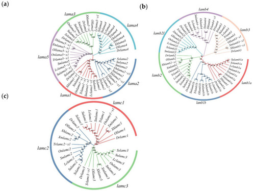
Phylogenetic analysis of laminin nucleotide sequence of ten species. Dr (Danio rerio), Lc (Larimichthys crocea), Sm (Scophthalmus maximus), Ol (Oryzias latipes), Tr (Takifugu rubripes), On (Oreochromis niloticus), Xm (Xiphophorus maculatus), Xh (Xiphophorus helleri), Ss (S. schlegelii) and Su (S. umbrosus). The numbers on the graph represent approval ratings. (a) Phylogenetic tree of laminin α chains; (b) Phylogenetic tree of laminin β chains; (c) Phylogenetic tree of laminin γ chains. The same gene from different species comes together to form a single branch with the same color. |
|
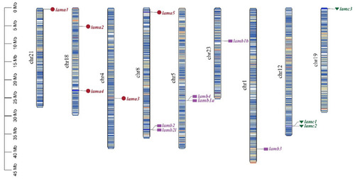
Chromosome location of laminin genes in S. schlegelii. Genes of the same subfamily are marked by the same color and shade. Chr: chromosome. |
|
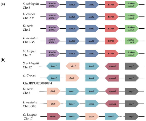
The position of tandem laminin genes in teleosts. The arrow direction indicates the direction of transcription. Dashed lines indicate discontinuities between genes. Genes of the same gene or family are represented in the same color. (a) Orders of lamb2 and lamb2l in teleosts; (b) Orders of lamc1 and lamc2 in teleosts. |
|
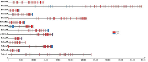
Gene structures of 14 laminin genes. The black solid line represents the intron region. The blue rectangle represents the UTR region, and the red rectangle represents the exon region. |
|
Conserved domains analysis of laminin genes. The number represents the length of the amino acid. Different colors of rectangle represent diverse domains. |
|
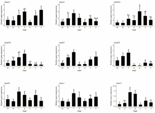
Expression analysis of laminin genes at seven different ovarian development stages. The relative expression levels of laminin genes are shown in different colors. III: ovary at stage III; IV: ovary at stage IV; V: ovary at stage V; B: blastula stage; G: gastrula stage; S: somites stage; P: prehatching stage. Different letters are signs of significant differences. |
|
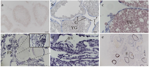
The location of lama4 in ovaries during oogenesis and gestation. (a) ovary at stage III; (b) ovary at stage IV—YG: yolk globule; T: theca cell; G: granulosa cell; (c) ovary at stage V; (d) embryos at the blastula stage—B: blastula stage, FP: follicular pseudoplacenta; (d’) follicular pseudoplacenta at the blastula stage—BV: blood vessel; (e) embryos at the prehatching stage—E: embryo. scale bar: 50 μm. |

