
- Title
-
Ethyl Acetate Fraction of Hedyotis diffusa Willd Induces Apoptosis via JNK/Nur77 Pathway in Hepatocellular Carcinoma Cells
- Authors
- Ning, W., Xu, N., Zhou, C., Zou, L., Quan, J., Yang, H., Lu, Z., Cao, H., Liu, J.
- Source
- Full text @ Evid. Based Complement. Alternat. Med.

Toxicity evaluation of HDW in zebrafish. 2 dpf larvae were randomly divided into 6 groups (n = 30) and immersed with HHDW (a), NHDW (b), EHDW (c), and PHDW (d), respectively. Mortality and malformation were monitored after 72 h. |
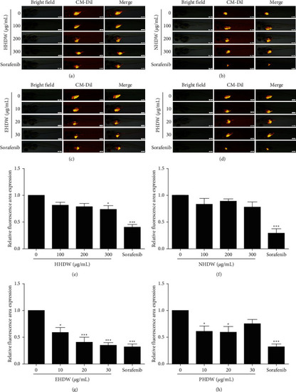
Antitumor activity of different extracts of HDW in zebrafish xenograft model. EHDW inhibited the proliferation of Hep3B cells in vivo. Hep3B cell-bearing zebrafish were treated with HHDW (a), NHDW (b), EHDW (c), PHDW (d), or sorafenib (0.25 μM) for 72 h and then CM-Dil-labeled Hep3B cells in yolk sac were visualized using a fluorescence microscope. Left: bright field, middle: CM-Dil-staining cells, right: merge. Bar: 200 μm. (e–h). Quantitative analysis of the fluorescent area in zebrafish. ∗P < 0.05, ∗∗P < 0.01, and ∗∗∗P < 0.001 vs. control. |
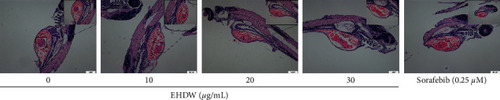
EHDW inhibited the tumorigenicity of Hep3B cells in zebrafish. Larvae were microinjected with 200 cells, and then treated with EHDW at 10, 20, and 30 μg/mL or sorafenib (0.25 μM). After 72 h, zebrafish were dehydrated and embedded in paraffin for HE staining. Bar: 50 μm. Red circles indicated tumor cells. |

EHDW exerted cytotoxicities in Hep3B cells in dose- and time-dependent manners. Hep3B cells were treated with various concentrations of EHDW for 24 and 48 h MTT assay was used to evaluate cell viability. |
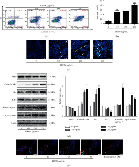
EHDW induced apoptosis in Hep3B cells. (a) Cell apoptosis was measured using flow cytometry. Representative data were analyzed with Annexin-V/PI staining in Hep3B cells after 24 h of exposure to indicated concentrations of EHDW. (b) Flow cytometry analysis of the percentage of apoptotic cells (early apoptosis and late apoptosis) from three independent experiments. ∗∗P < 0.01 and ∗∗∗P < 0.001 vs. control. (c) Apoptotic morphological observation. Fluorescence microscope images showed morphological changes in Hep3B cells upon 150–250 μg/mL EHDW treatment for 24 h White arrows showing bright blue regions indicated nuclear pyknosis and fragmentation of chromatin. (d) Expression of cyto c in cytosol and PARP, cleaved-PARP, Bcl-2, BAX, cleaved-caspase 3 in total proteins were examined by Western blotting. ∗P < 0.05, ∗∗P < 0.01, and ∗∗∗P < 0.001 vs. control. One-way ANOVA, post hoc comparisons, Tukey's test. Columns, means, error bars, SEM. (e) EHDW induced apoptosis in vivo. The 5-μm zebrafish tissue sections were deparaffinized and stained with Hoechst 33258 dye. Representative fluorescent photographs were taken by fluorescence microscopy. |
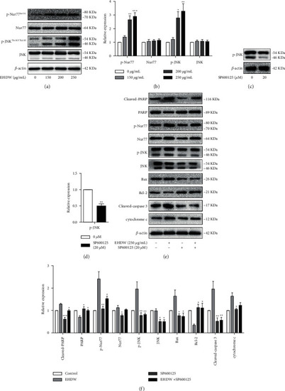
EHDW induced apoptosis in Hep3B cells by JNK/Nur77 pathway. (a–b) EHDW activated JNK/Nur77 pathway. After incubation for 24 h, the protein levels of JNK, p-JNK(Thr183/Tyr185), Nur77, and p-Nur77(Ser351) were measured by Western blotting. ∗P < 0.05, ∗∗P < 0.01, and ∗∗∗P < 0.001 vs. control. One-way ANOVA, post hoc comparisons, Tukey's test. Columns, means, error bars, SEM. (c–d) The functional verification of SP600125. Hep3B cells were treated with SP600125 for 24 h Western blotting was performed to detect the expression of p-JNK. ∗P < 0.05, ∗∗P < 0.01, and ∗∗∗P < 0.001 vs. control. One-way ANOVA, post hoc comparisons, Tukey's test. Columns, means, error bars, SEM. (e–f) The apoptotic effect of EHDW were counteracted in the presence of SP600125. The expression of proteins in JNK/Nur77 pathway and the downstream-related proteins were detected after the cells were treated with SP600125 and EHDW for 24 h. ∗P < 0.05, ∗∗P < 0.01, and ∗∗∗P < 0.001 vs. EHDW treatment. One-way ANOVA, post hoc comparisons, Tukey's test. Columns, means, error bars, and SEM. |