
|
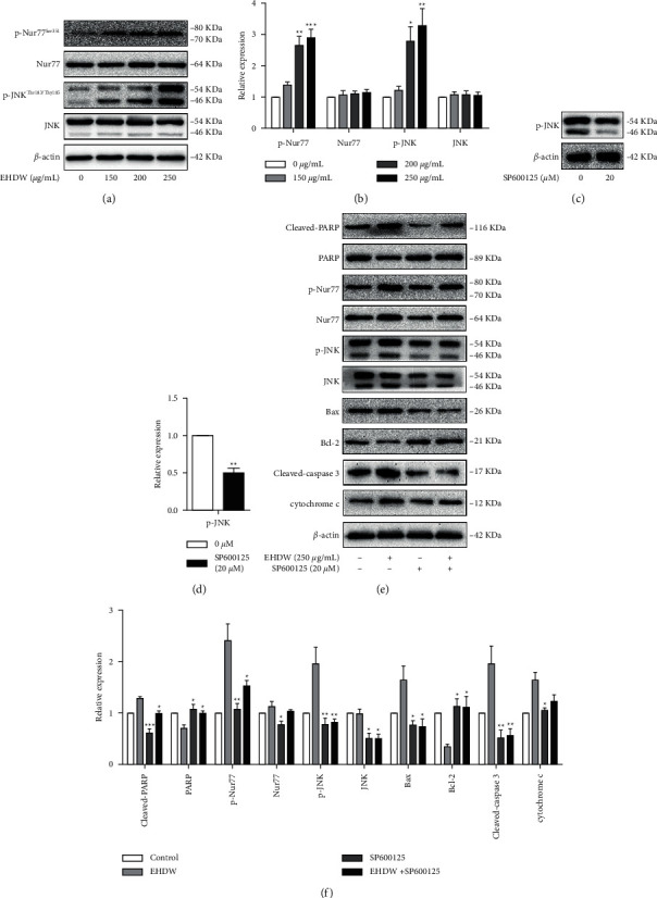
Fig. 6
EHDW induced apoptosis in Hep3B cells by JNK/Nur77 pathway. (a–b) EHDW activated JNK/Nur77 pathway. After incubation for 24 h, the protein levels of JNK, p-JNK(Thr183/Tyr185), Nur77, and p-Nur77(Ser351) were measured by Western blotting. ∗P < 0.05, ∗∗P < 0.01, and ∗∗∗P < 0.001 vs. control. One-way ANOVA, post hoc comparisons, Tukey's test. Columns, means, error bars, SEM. (c–d) The functional verification of SP600125. Hep3B cells were treated with SP600125 for 24 h Western blotting was performed to detect the expression of p-JNK. ∗P < 0.05, ∗∗P < 0.01, and ∗∗∗P < 0.001 vs. control. One-way ANOVA, post hoc comparisons, Tukey's test. Columns, means, error bars, SEM. (e–f) The apoptotic effect of EHDW were counteracted in the presence of SP600125. The expression of proteins in JNK/Nur77 pathway and the downstream-related proteins were detected after the cells were treated with SP600125 and EHDW for 24 h. ∗P < 0.05, ∗∗P < 0.01, and ∗∗∗P < 0.001 vs. EHDW treatment. One-way ANOVA, post hoc comparisons, Tukey's test. Columns, means, error bars, and SEM.