
- Title
-
Estrogens revert neutrophil hyperplasia by inhibiting Hif1α-cMyb pathway in zebrafish myelodysplastic syndromes models
- Authors
- Li, X., Wang, L., Qin, X., Chen, X., Li, L., Huang, Z., Zhang, W., Liu, W.
- Source
- Full text @ Cell Death Discov

Estrogens decrease neutrophils in c-mybhyper transgenic zebrafish.
A Flowchart for drug screening in c-mybhyper transgenic zebrafish. For preliminary screening, 96-well plates were used and each well was placed 5 embryos to detect potential effective drugs. For secondary screening, 12-well plates were used and each well was placed more embryos (n > 15) to further verify the potential effective drugs screened from the preliminary screening. B Natural estrogens decreased SB positive cells in the CHT region in c-mybhyper transgenic zebrafish. Many pairs of zebrafish parents were selected for the drugs treatment, the embryos from each parent pair were randomly divided into two groups, one for a compound treated group and the other for its control (DMSO group). (t-test, ***p < 0.001, **p < 0.01. n > 15.) C Progesterone had no effect on SB positive cells in the CHT region in c-mybhyper transgenic zebrafish. (t-test, ns, no significance. n > 15). |

E2 decreases the number of neutrophils in c-mybhyper zebrafish, mainly through inhibiting cell proliferation and promoting cell apoptosis.
A E2 exposure decreased SB positive cells in the CHT region. (t-test, ***p < 0.001, **p < 0.01. n > 20). B E2 exposure decreased lyz in the CHT region, as determined by WISH. (t-test, ***p < 0.001, **p < 0.01. n > 20). C The qPCR quantification of the decrease in lyz expression with E2 (t-test, mean ± SEM; ***p < 0.001, **p < 0.01. n ≥ 10). D May-Grunwald-Giemsa staining of whole KM blood cells in 6-month-old c-mybhyper animals followed by four days of E2 treatment (t-test, ***p < 0.001. n = 12). Red arrowheads, blue asterisks, black arrowheads and yellow lightning indicate neutrophils, precursors, lymphocytes and macrophages, respectively. E Double staining of bromodeoxyuridine (BrdU)/Lcp indicated decreased neutrophil proliferation in c-mybhyper zebrafish embryos treated with E2. (one-way ANOVA (LSD) ***p < 0.001, n = 12). F The TUNEL assays showed the effect of E2 on the apoptosis of myeloid lineage in zebrafish embryos (one-way ANOVA (LSD) ***p < 0.001, ns, no significance. n = 12). |
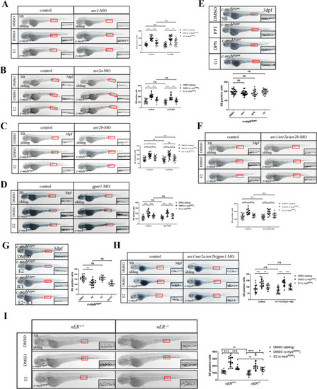
E2 decreases neutrophils in c-mybhyper transgenic zebrafish embryos through ERs-independent mechanisms.
A–D Knockdown of esr1 (A), esr2a (B), esr2b (C) and gper1 (D) did not alleviate E2-mediated inhibition of c-mybhyper zebrafish neutrophils. (one-way ANOVA (LSD) ***p < 0.001, **p < 0.01, *p < 0.05. ns, no significance, n > 15). E PPT, DPN and G1 had no effect on c-mybhyper zebrafish neutrophils. (one-way ANOVA (LSD), ns, no significance, n > 20). F Knockdown of esr1, esr2a and esr2b simultaneously did not alleviate E2-mediated inhibition of c-mybhyper zebrafish neutrophils. (one-way ANOVA (LSD) ***p < 0.001, **p < 0.01, ns, no significance, n > 15). G ICI 182780 did not alleviate E2-mediated inhibition of c-mybhyper zebrafish neutrophils. (one-way ANOVA (LSD) ***p < 0.001, ns, no significance, n > 15). H Knockdown of esr1, esr2a esr2b and gper1 simultaneously did not alleviate E2-mediated inhibition of c-mybhyper zebrafish neutrophils (one-way ANOVA (LSD) ***p < 0.001, **p < 0.01, ns, no significance, n > 15). I The triple esr1; esr2a; esr2b receptor mutant (esr-/-) was produced through mating to knock out all three classical nuclear receptors. SB staining showed that the E2-induced neutrophil decrease was also not alleviated in the triple mutant. (t-test, mean ± SEM; ***p < 0.001, *p < 0.05. ns, no significance. n > 10). |
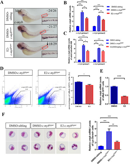
E2 decreases c-myb expression in c-mybhyper transgenic zebrafish embryos.
A E2 exposure decreased c-myb in the CHT, as determined by WISH. B qPCR quantification of the decreased c-myb expression by E2 (the c-mybhyper fusion gene contained two parts, which comprised truncated c-myb from exon1 to exon10, followed by a near full-length c-myb from exon2 to exon 15). c-myb-primer1 and c-myb-primer2 were designed at repetitive and nonrepetitive sequences. (t-test, mean ± SEM. ***p < 0.001, **p < 0.01, n > 10). C qPCR quantification of decreased c-myb expression in c-mybhyper adult zebrafish kidney after treatment with E2. (t-test, mean ± SEM. ***p < 0.001, *p < 0.05, n > 10) (D) FACS analysis confirmed that E2 diminished c-myb:GFP+ cells. (t-test, mean ± SEM. *p < 0.05, n > 10) (E) qPCR quantification of decreased c-myb expression in c-myb:GFP+ cells by E2. (t-test, mean ± SEM. ***p < 0.001, n > 10). F qPCR quantification of decreased c-myb expression in lyz:Dsred+ cells by E2. (t-test, mean ± SEM. ***p < 0.001, **p < 0.01, n > 10). |

Hif1α participates in E2 induced down-regulation of c-myb and neutrophil hyperplasia.
A Double staining of hif1α and c-myb-GFP with antibodies with or without E2 treatment. B Knockdown (MO) or inhibition (PX-478) of hif1α decreased c-myb and lyz expression by WISH as well as SB positive neutrophils in c-mybhyper zebrafish. (t-test, ***p < 0.001, **p < 0.01, *p < 0.05, n > 20). C Overexpression of hif1α reversed E2 induced down-regulation of c-myb and lyz expression by WISH and neutrophil hyperplasia by SB staining. (t-test, ***p < 0.001, **p < 0.01, *p < 0.05, n > 20). |

E2 enhances neutrophil apoptosis through suppressing the expression of hif1α and c-myb under physiological conditions.
A E2 decreased neutrophils in AB zebrafish embryos. (t-test, ***p < 0.001, n > 20). B May–Grunwald–Giemsa staining of whole KM blood cells in 6-month-old AB zebrafish after 4 days of E2 treatment (t-test, ***p < 0.001, n = 12). Red arrowheads, blue asterisks, black arrowheads and yellow lightning indicates neutrophils, precursors, lymphocytes and macrophages, respectively. C Staining of hif1α with antibody with or without E2 treatment. D E2 exposure decreased c-myb in the CHT, as determined by WISH. E qPCR quantification of decreased c-myb expression in lyz:Dsred+ cells by E2 (t-test, mean ± SEM. ***p < 0.001, **p < 0.01, *p < 0.05, n > 20). F Effect of E2 on neutrophil proliferation in AB zebrafish embryos. (one-way ANOVA (LSD). ns, no significance, n > 10). G E2 promotes the apoptosis of myeloid lineage cells in AB zebrafish embryos (one-way ANOVA (LSD) **p < 0.01. n > 10). |
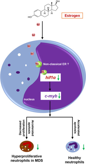
Schematic illustration of the neutrophil hyperplasia inhibiting role of E2 in zebrafish MDS E2 reverts neutrophil hyperplasia by regulating the proliferation and apoptosis of neutrophils through inhibition of the hif1α-c-myb pathway in zebrafish MDS models. |