
- Title
-
Reintroduction of DJ-1 in Müller Cells Inhibits Retinal Degeneration in the DJ-1 Deficient Retina
- Authors
- Gharbi, N., Røise, D., Førre, J.E., Edson, A.J., Hushagen, H.A., Tronci, V., Frøyset, A.K., Fladmark, K.E.
- Source
- Full text @ Antioxidants (Basel)
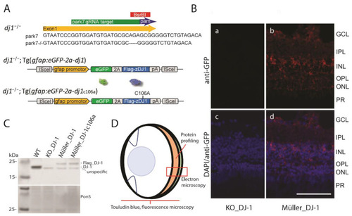
Zebrafish lines and workflow. (A) A DJ-1 knockout line was previously established by using the CRISPR-cas9 method to target a 20 bp region of exon one of the park7 gene [19]. Lines expressing glial specific wild-type DJ-1 or DJ-1 c106a in a DJ-1 null background were constructed by using ISce1 transgenesis and regulatory elements of glial fibrillary acidic protein (GFAP). The viral 2A peptide allows expression of GFP and Flag-DJ1 as uncoupled protein. In the retina, the gfap promotor drives expression only in the Müller glia cells. (B) Glial expression of GFP in the Müller_DJ-1 line (b,d) versus KO_DJ-1 (a,c). Note that GFP expression was determined by using a GFP antibody, followed by a far-red secondary antibody to avoid interference from retinal autofluorescence. Prominent expression is found around the Müller cell bodies. GFP expression also extends through the Müller processes into the photoreceptor layer. Faint expression can be observed in the Müller foot processes on the inner limiting membrane. Bar, 50 μm. (C) A Western blot shows expression of endogenous DJ-1 and Flag-tagged DJ-1 from total brain extracts belonging to animals from which eyes were collected. Ponceau S staining was used as a loading control. Asterisk points to an unspecific band. (D) Sagittal view of the zebrafish eye and workflow employed in this study. |
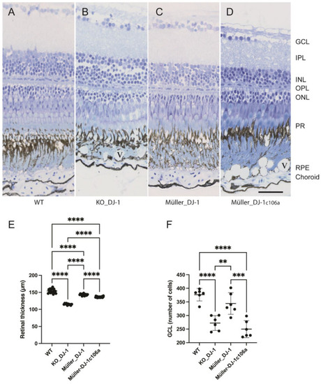
Laminar morphology of wild-type, knockout and transgenic retinas. Light microscopic images of toluidine-blue-stained retinal cross-sections from nine-month-old adult zebrafish: (A) wild type, (B) DJ-1 knockout, (C) Müller-cell-expressed wild-type DJ-1, (D) Müller-cell-expressed DJ-1c106a mutant. (E). Retinal thickness (μm) measured on eye sections from three fish in each group. **** p < 0.0001, one-way ANOVA, n = 17 (WT), 10 (KO_DJ-1), 15 (Müller_DJ-1) and 19 (Müller_DJ-1c106a). (F) Number of cells in ganglion cell layer measured on sections from three fish; ** p < 0.01, *** p < 0.001 and **** p < 0.0001 versus wild type, one-way ANOVA, n = 6). GCL, ganglion cell layer; IPL, inner plexiform layer; INL, inner nuclear layer; OPL, outer nuclear layer; PR, photoreceptors; RPE, retinal pigment epithelium; V, vacuole in RPE layer. Bar, 20 μm applies to all panels. |
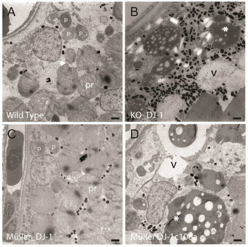
Ultrastructural changes in the retinal pigment epithelium induced by the loss of DJ-1 are inhibited by introducing DJ-1 in Müller cells. Electron micrographs of photoreceptors (pr) and retinal pigment epithelium (RPE) of wild-type (A), DJ-1 knockout (B), Müller-expressed DJ-1 (C) and Müller-expressed DJ-1c106a mutant (D) retinas from 4-month-old adult zebrafish. DJ-1-deficient retinas displayed a high degree of vacuolization (v), in addition to large electron-dense structures (*) in the RPE. The large electron-dense structures contained remnants of membranous/filamentous structures. Transgenic retinas expressing Müller-selective DJ-1c106a mutant displayed similar morphology as DJ-1 knockouts. The ultrastructural changes induced by DJ-1 loss were prevented by Müller cell selective expression of wild type DJ-1. Different stages of phagosomes containing segment filaments can be seen in wild-type and Müller_DJ-1 retinas (marked with arrowhead and p). Bar, 1 µm. PHENOTYPE:
|
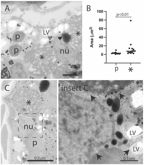
Ultrastructural features in the retinal pigment epithelium of the DJ-1-deficient retina. Electron micrographs of the RPE area of DJ-1 knockouts (A,C). Images show both phagosomes (p) and the larger electron-dense structures (*), in addition to numerous small electron lucent vacuoles (LV) in RPE. Phagosomes and electron lucent vacuoles were surrounded by membranes (arrows), but no clear limited membrane was observed limiting the electron-dense structures. (B) Quantitation of area of phagosomes (p) and electron-dense structures (*); p < 0.01. Mann-Whitney-test, n = 12 (WT) and 15 (KO_DJ-1); p, phagosome; * electron-dense structure; LV, electron-lucent vacuole; nu, RPE nucleus. PHENOTYPE:
|