
- Title
-
A Novel Pseudoalteromonas xiamenensis Marine Isolate as a Potential Probiotic: Anti-Inflammatory and Innate Immune Modulatory Effects against Thermal and Pathogenic Stresses
- Authors
- Wasana, W.P., Senevirathne, A., Nikapitiya, C., Eom, T.Y., Lee, Y., Lee, J.S., Kang, D.H., Oh, C., De Zoysa, M.
- Source
- Full text @ Mar. Drugs
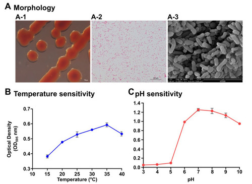
Morphological and physiological characterization of Pseudoalteromonas xiamenensis S1131. (A) Colony morphology on marine agar (A-1), Gram staining (A-2), and scanning electron microscopy (A-3) demonstrating the morphological appearance of the P. xiamenensis isolate. Scale bars indicate 100 µm, 10 µm, and 5 µm length for colony morphology, Gram staining, and SEM, respectively. Physiological characterization determining the optimum temperature (B) and pH (C) conditions. Cultures were incubated for 24 h under specified temperatures and pH conditions and the absorbance measurements were taken at OD595. |
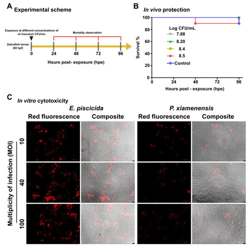
Safety assessments of the probiotic isolate, P. xiamenensis, and pathogenic strain, E. piscicida. In vivo safety assessment was conducted by exposing zebrafish larvae to different concentrations of P. xiamenensis in egg water. (A) Schematic representation for the experimental schedule of P. xiamenensis exposure to the zebrafish larvae. (B) Kaplan−Meier survival curves of zebrafish larvae upon exposure to the different concentrations of P. xiamenensis. (C) In vitro cytotoxicity assay. Fathead minnow (FHM) fish epithelial cells were either exposed to P. xiamenensis probiotic isolate, or E. piscicida pathogenic strain. After 1 h of incubation, cells were washed with PBS and non-internalized bacteria were eliminated by gentamycin treatment. The cells were replenished with complete growth media and further incubated for 12 h. After 12 h incubation, the cells were subjected to propidium iodide (PI) staining and observed under a fluorescent microscope. Images were taken at 200 × magnification. |
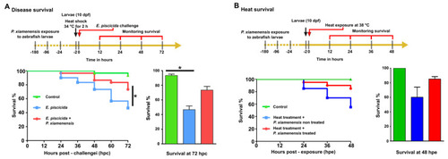
Effect of P. xiamenensis on disease resistance and heat stress survival. (A) Treatment with P. xiamenensis was evaluated for disease resistance enhancement against fish-pathogenic E. piscicida. After P. xiamenensis treatment, zebrafish larvae were challenged with E. piscicida. Mortality was recorded for 72 h post-exposure (hpe) and the relative percent survival was determined. Asterisk (*) indicates a significant difference in comparison to the non-infected control. (B) Enhancement of heat resistance was evaluated for zebrafish after P. xiamenensis treatment at 28 °C followed by heat stress exposure at 38 °C for 48 h. The mortality of fish larvae was evaluated. Statistical significance was set at p < 0.05. |

qRT-PCR and immunoblotting analysis of P. xiamenensis-treated zebrafish larvae. (A) The effect of P. xiamenensis treatment on immune-related genes at the mRNA level using qRT-PCR. Gene expression profiles were determined in non-treated control and P. xiamenensis-treated zebrafish treatment groups. Asterisk (*) indicates a significant difference compared to the non-treated control. Significance was set at p < 0.05. (B) Immunoblot analysis of Hsp90 and Tnfα in P. xiamenensis-treated zebrafish larvae, compared to the non-treated control. Expression levels of β-actin were set as the normalizing control. The intensity of each protein band was measured by Image J software. Asterisk (*) indicates a significant difference compared to the control group (* p < 0.05, ** p < 0.01). Statistical significance was set at p < 0.05. |

Mucin-dependent colony dispersion and macrophage uptake using motile pathogens S. typhimurium and E. piscicida. (A) The effect of mucin on colony dispersion/prevention of biofilms was investigated using the motile bacterium S. typhimurium and E. piscicida. Porcine mucin at variable concentrations was incorporated into soft agar and allowed to solidify. Mid-log phase bacterial culture was spotted on the agar. After 12 h incubation at 28 °C for E. piscicida and 37 °C for S. typhimurium, the colony diameter was measured. ** and *** indicate a significant difference compared to the non-treated control. (B) The mouse macrophage RAW264.7 cell line was seeded in 24-well plates at 5 × 10⁴ cells/well. Cell media was supplemented with porcine mucin at variable concentrations. S. typhimurium and E. piscicida were inoculated at MOI 40 and 100. After 1 h of co-incubation at 37 °C, the cells were washed with PBS three times, treated with gentamycin, and lysed with 0.05% Triton X-100 PBS. The whole lysate was collected and serially diluted for plating on agar. Internalized bacterial numbers were determined by CFU counting. The relative uptake was calculated based on the uptake of the control. ** and *** indicate a significant difference compared to the non-treated control. The level of significance was set at p < 0.05. |