FIGURE SUMMARY
Title

Hearing loss genes reveal patterns of adaptive evolution at the coding and non-coding levels in mammals

Authors
Trigila, A.P., Pisciottano, F., Franchini, L.F.
Source
Full text @ BMC Biol.

Bioinformatics pipeline and filtering process of two datasets of hearing loss genes: the classic dataset composed of non-syndromic hearing loss genes identified in humans which is composed of a total of 129 coding genes obtained by combining the information available in the “Hereditary Hearing Loss Homepage” [17], the “Genetics Home Reference – Nonsyndromic deafness related genes” website [18], the Connexin-deafness homepage – Nonsyndromic deafness related connexins” [19] and from the chapter “Deafness and Hereditary Hearing Loss Overview” from the online book “Gene Review” [20], and the recently identified hearing impairment genes by the International Mouse Phenotype Consortium [2123]

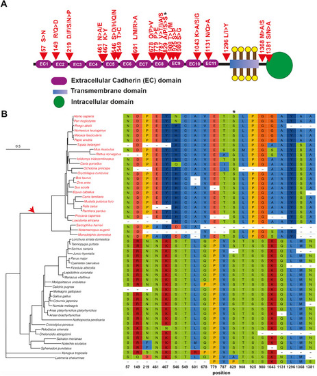
Phylogenetic tree and positive selected sites of an essential tip link protein: PCDH15. A Schematic diagram of the PCDH15 protein domains showing the approximate localization of positively selected sites (red arrows). Protein domains were approximately depicted according to UniProtKB - A9Z1W1 (A9Z1W1_HUMAN). B On the left: phylogenetic tree showing vertebrate species used for the analysis. The red arrow indicates the foreground branch. Mammalian species included in the foreground branch are in red. On the right: sequence alignment of positively selected sites identified. Positions of the human sequence given correspond to the ENST00000373965.6 transcript. Note that a change of Serine by Serine (asterisk) is identified as a positive selected site: serine is the only amino acid that is encoded by two disjoint codon sets so that a tandem substitution of two nucleotides is required to switch between the two sets. It has been suggested that the great majority of codon set switches proceed by two consecutive nucleotide substitutions, via a threonine or cysteine intermediates, are driven by selection even though this may not be the kind of positive selection driving functional divergences [29, 30]

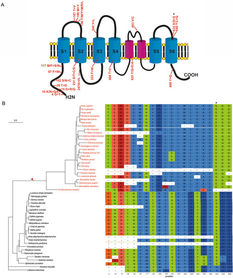
Phylogenetic tree and positive selected sites of an essential tip link protein: TMC1. A Schematic diagram of the TMC1 protein domains (taken from [40]) showing the approximate localization of positively selected sites. Transmembrane domains (S1 to S6) were depicted using info from UniProtKB - Q8TDI8 (TMC1_HUMAN). B On the left: phylogenetic tree showing vertebrate species used for the analysis. The red arrow indicates the foreground branch. Mammalian species included in the foreground branch are written in red. On the right: sequence alignment of the positively selected sites identified. Positions of the human sequence given correspond to the ENST00000645208.2 transcript. Note that a change of Serine by Serine (asterisk) is identified as a positive selected site, see Fig. 2 legend for an explanation

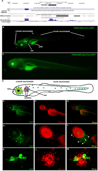
Representative transgenic zebrafish carrying the TSAR4204-JAZF1. A Location of the TSAR.4204 in the JAZF1 locus in chromosome 7 of the human genome (modified from UCSC Genome Browser). B Low-magnification microphotograph showing the expression pattern of the transgene TSAR.4204-cFos-eGFP (human sequence as a mammalian representative) in a transgenic zebrafish at 6 dpf. C Low-magnification microphotograph showing the expression pattern of the transgene TSAR.4204-Gg-cFos-eGFP carrying the chicken ortholog of the TSAR.4204 sequence in a transgenic zebrafish at 6 dpf. D Scheme of a 6 dpf zebrafish showing the approximate location of the inner ear and neuromasts of the lateral line, where the transgene carrying the mammalian sequence of TSAR.4204 directs the expression of eGFP. Figures (E–M) show magnifications of the head region of a transgenic zebrafish carrying the human sequence of TSAR.4204-cFos-eGFP exhibiting in major detail the expression of eGFP (E, H, K), the fluorescent marker FM4-64 that specifically labels hair cells (F, I, L), and a merged image showing the colocalization of eGFP and the hair cell marker (G, J, M). Figures (K–M) show in detail confocal images of neuromast expressing eGFP in the lateral line of transgenic zebrafish carrying the human sequence of TSAR.4204-cFos-eGFP. We show the best representative image for each line

Key genes involved in the mechanotransduction machinery display signatures of positive selection. A A network interaction map of all the 53 positively selected proteins from the STRING database (https://string-db.org). Edges represent protein associations, with either known or predicted interactions. Known interactions are represented by light-blue lines (curated databases) or purple lines (experimentally determined). Predicted interactions are represented by green (neighbors), red (fusions), and blue (co-occurrence) edges. Other interactions are represented by gold (text-mining), black (co-expression), and gray (protein-homology) lines. Interaction is observed between classical, well-known hearing genes (PCDH15, CDH23, TMC1) related to the mechanotransduction machinery and novel genes (LOXHD1), for which its role in this complex has yet to be explored but still show strong signals of positive selection. Genes involved in the tip link function are highlighted in red (B). Cellular organization of the mammalian hair cell. Genes that play key roles in hair cell function and displayed signatures of positive selection in mammals are highlighted in red. C Tip link schematic showing the location of key genes involved in function in this particular structure. Genes involved in the mechanotransduction machinery that display signatures of positive selection are highlighted in red

Acknowledgments
This image is the copyrighted work of the attributed author or publisher, and ZFIN has permission only to display this image to its users. Additional permissions should be obtained from the applicable author or publisher of the image. Full text @ BMC Biol.