FIGURE SUMMARY
Title

Recurrent NFIA K125E substitution represents a loss-of-function allele: Sensitive in vitro and in vivo assays for nontruncating alleles

Authors
Uehara, T., Sanuki, R., Ogura, Y., Yokoyama, A., Yoshida, T., Futagawa, H., Yoshihashi, H., Yamada, M., Suzuki, H., Takenouchi, T., Matsubara, K., Hirata, H., Kosaki, K., Takano-Shimizu, T.
Source
Full text @ Am. J. Med. Genet. A

Clinical characteristics of two patients with the same NFIA variant. (a) Results of head MRI of Patient 1 at 5 years of age. The picture above shows sagittal T1‐weighted image. The picture below shows axial T2‐weighted fluid‐attenuated inversion recovery image. Note thin corpus callosum, cyst of septi pellucidi, ventricular wall irregularity, and decreased white matter volume. (b) Pictures of Patient 1 at 6 years of age. Note the high hairline, small eyes, anteverted nares, a depressed nasal bridge, a broad columella, and a thin upper‐lip. (c) Results of head MRI of Patient 2 at 1 month of age. The picture above shows sagittal‐T2‐weighted image. The picture below shows axial diffusion‐weighted image. Note polycerebral gyrus at parasylvius fissures, cortical dysplasia of bilateral cerebral hemisphere, partial myelination delay, and hypoplasia of corpus callosum [Color figure can be viewed at wileyonlinelibrary.com]

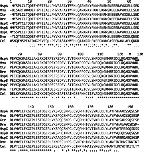
Amino acid sequence alignment of the DNA-binding domains of human NFIA with its orthologs and paralogs. Sequences and domain definitions were obtained from UniProt (The UniProt Consortium, 2019). HspA: human NFIA (Q12857, residues 1–194); HspB: human NFIB (O00712, 1–195); HspC: human NFIC (P08651, 1–195); HspX: human NFIX (Q14938, 1–194); Mmu: mouse Nfia (Q02780, 24–217); Xtr: frog nfia (A0A6I8RQ42, 1–194); Dre: zebrafish nfia (F1R2R8, 1–193); Dme: fly NfI (Q86P06, 19–212); and Cel: nematode nfi-1 (Q09631, 44–237). The K125E mutation site is indicated by an arrow; 6th and 7th mutagenized sites in Armentero et al. (1994) are underlined

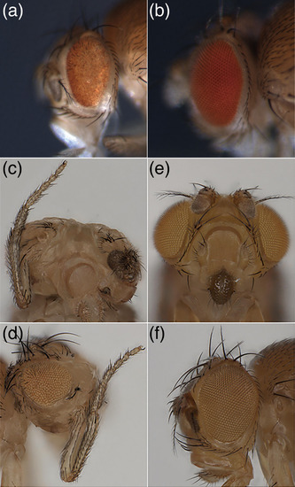
Phenotypes of ectopic expression of
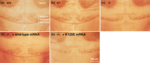
 NFIA gene in Drosophila. Ectopic expression of NFIAWT (a) but not NFIAK125E (b) during retinal development (GMR>NFIAWT) caused severe neurodegeneration (rough eye phenotype). Ectopic expression of NFIAWT (c and d) but not NFIAK125E (e and f) under the control of the ey‐GAL4 driver caused antenna‐to‐leg transformation. Note that the ey gene is expressed in the eye‐antennal disc primordia and ubiquitously in the first instar larval disc (Quiring et al., 1994; Urbach & Technau, 2003) [Color figure can be viewed at wileyonlinelibrary.com]

Commissural defects in nfia mutant zebrafish. Commissural axons (arrows) cross the midline in the midbrain/hindbrain boundary in wild‐type (a) (n = 4/4) and nfia heterozygous mutant (b) (n = 6/6) embryos at 3 dpf, but many of them failed to do so in nfia homozygous mutants (c) (n = 5/5). The midline crossing defects in nfia homozygous mutants were rescued by injection of NFIAWT mRNA (d) (n = 5/5), but not by that of NFIAK125E one (e) (n = 5/5). Axons were labeled with anti‐acetylated α‐tubulin antibody [Color figure can be viewed at wileyonlinelibrary.com]

HES1 and GFAP promoter assay in HEK293T cells. (a) Schematic representation of the human HES1 promoter‐NlucP and the human GFAP promoter‐NlucP constructs. Potential NFI‐binding sites (NNTTGGCNNNNNNCCNNN) predicted by TFBIND (http://tfbind.hgc.jp/; Tsunoda & Takagi, 1999) are shown by triangles. (b) HES1 promoter repression by NFIA. Dose‐dependent repression of human HES1 promoter by NFIAWT was not observed in NFIAK125E. Data are mean ± SD from triplicate experiments. **p < 0.01 by lower‐tailed Dunnett multiple comparisons test (α = 0.05). ns, not significant. (c) GFAP promoter activation by NFIA. Dose‐dependent activation of human GFAP promoter by NFIAWT was weakened in NFIAK125E. Data are mean ± SD from triplicate experiments. **p < 0.01 by upper‐tailed Dunnett multiple comparisons test (α = 0.05). ##p < 0.01 by unpaired t‐test. ns, not significant

Acknowledgments
This image is the copyrighted work of the attributed author or publisher, and ZFIN has permission only to display this image to its users. Additional permissions should be obtained from the applicable author or publisher of the image. Full text @ Am. J. Med. Genet. A