IMAGE

FIGURE 1

ID
ZDB-IMAGE-210708-86
Source
Figures for Uehara et al., 2021
Image
Figure Caption

FIGURE 1

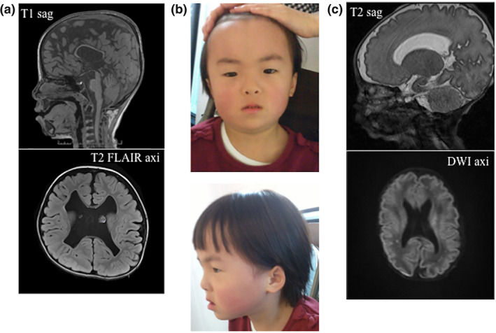
Clinical characteristics of two patients with the same NFIA variant. (a) Results of head MRI of Patient 1 at 5 years of age. The picture above shows sagittal T1‐weighted image. The picture below shows axial T2‐weighted fluid‐attenuated inversion recovery image. Note thin corpus callosum, cyst of septi pellucidi, ventricular wall irregularity, and decreased white matter volume. (b) Pictures of Patient 1 at 6 years of age. Note the high hairline, small eyes, anteverted nares, a depressed nasal bridge, a broad columella, and a thin upper‐lip. (c) Results of head MRI of Patient 2 at 1 month of age. The picture above shows sagittal‐T2‐weighted image. The picture below shows axial diffusion‐weighted image. Note polycerebral gyrus at parasylvius fissures, cortical dysplasia of bilateral cerebral hemisphere, partial myelination delay, and hypoplasia of corpus callosum [Color figure can be viewed at wileyonlinelibrary.com]

Acknowledgments
This image is the copyrighted work of the attributed author or publisher, and ZFIN has permission only to display this image to its users. Additional permissions should be obtained from the applicable author or publisher of the image. Full text @ Am. J. Med. Genet. A