
- Title
-
Caffeine-Cyclodextrin Complexes as Solids: Synthesis, Biological and Physicochemical Characterization
- Authors
- Szmeja, S., Gubica, T., Ostrowski, A., Zalewska, A., Szeleszczuk, Ł., Zawada, K., Zielińska-Pisklak, M., Skowronek, K., Wiweger, M.
- Source
- Full text @ Int. J. Mol. Sci.
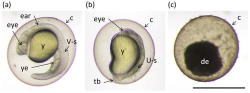
Examples of different phenotypes of zebrafish embryos at 24 h post fertilization (hpf) found in the bioassay described in the Section 2.6 “Toxicity”. (a) Normally developed embryo; (b) embryo with underdeveloped eye, brain and impaired extensions of yolk sack and tail; and (c) dead embryo. Ear; eye; y: yolk; ye: yolk extension; V-s: v-shaped somite; U-s: abnormally-shaped somite; tb: tail bud; and de: decomposing embryo. Scale bar equals 1 mm. |
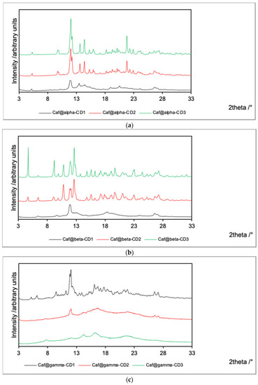
PXRD patterns for Caf@CD obtained by three methods (1: cogrinding; 2: cogrinding with the addition of water drops; and 3: depositing from soln.). (a) α-CD; (b) β-CD; and (c) γ-CD series. |
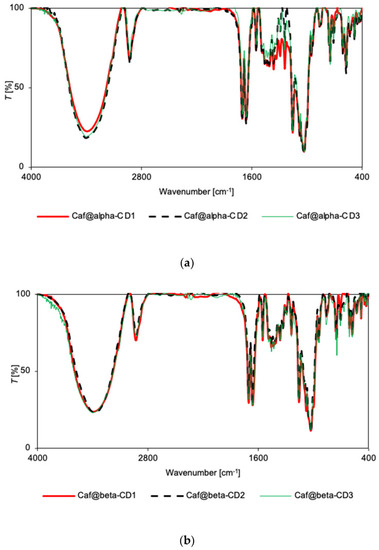
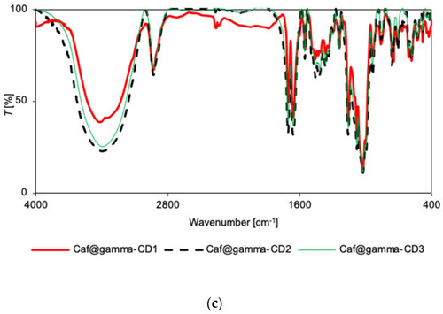
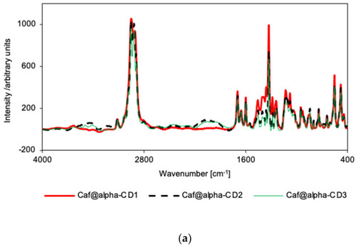
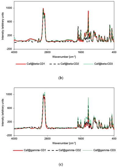
FT-IR spectra for Caf@CD obtained by three methods (1: cogrinding; 2: cogrinding with the addition of water drops; and 3: depositing from soln.). (a) α-CD; (b) β-CD; and (c) γ-CD series. |
FT-IR spectra for Caf@CD obtained by three methods (1: cogrinding; 2: cogrinding with the addition of water drops; and 3: depositing from soln.). (a) α-CD; (b) β-CD; and (c) γ-CD series. |
Raman spectra for Caf@CD obtained by three methods (1: cogrinding; 2: cogrinding with the addition of water drops; and 3: depositing from soln.). (a) α-CD; (b) β-CD; and (c) γ-CD series. |
Raman spectra for Caf@CD obtained by three methods (1: cogrinding; 2: cogrinding with the addition of water drops; and 3: depositing from soln.). (a) α-CD; (b) β-CD; and (c) γ-CD series. |
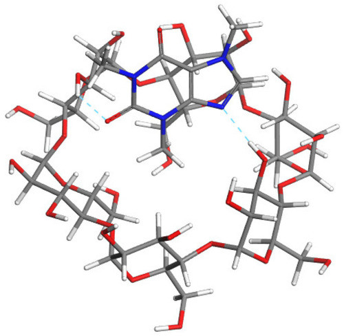
The most stable modeled structures: Caf@α-CD (upper); Caf@β-CD (middle); and Caf@γ-CD (lower). Atoms: C (grey); H (white); O (red); and N (blue). The cyan and pink dashed lines represent possible hydrogen bonds and close contacts stabilizing the complexes, respectively. |
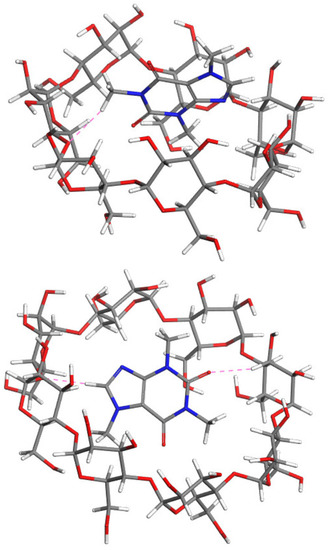
The most stable modeled structures: Caf@α-CD (upper); Caf@β-CD (middle); and Caf@γ-CD (lower). Atoms: C (grey); H (white); O (red); and N (blue). The cyan and pink dashed lines represent possible hydrogen bonds and close contacts stabilizing the complexes, respectively. |
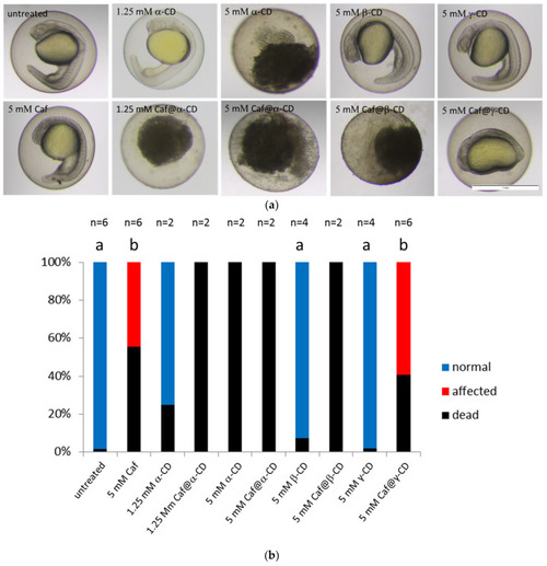
The effect of Caf@CDs on early development of the zebrafish embryo. (a) Images of zebrafish embryos after exposure to caffeine (Caf), CDs or Caf@CDs from 4 till 24 hpf. Embryos exposed to Caf@γ-CD show underdeveloped eyes, ears and severely impaired convergent extension. Less severe morphological abnormalities were also present in embryos exposed to 5 mM Caf. Exposure to 5 mM α-CD, 1.25 mM Caf@α-CD, or 5 mM Caf@β-CD was lethal to the embryos. Examples of live embryos at a most advanced developmental stage are shown. Scale bar, 1 mm. (b) Changes in the proportion between the dead, morphologically abnormal (affected), and unaffected (normal) embryos. The percentage of embryos which died upon treatment with Caf or Caf@CDs was significantly higher than that of untreated control. Also, pure α-CD was more toxic than the other two CDs, whereas β-CD and γ-CD had no effect on both the fish survival and their morphology. In order to reduce the number of animals, in the case of treatments which caused 100% rapid mortality, the experiments were repeated twice. Bars present mean values, n is the number of repetitions, each on 24 embryos. Mann-Whitney U test was performed on samples with ≥3 biological replicas. Statistically significant differences (p < 0.01) were marked with different letters. |