
- Title
-
Developmental and Neurotoxicity of Acrylamide to Zebrafish
- Authors
- Park, J.S., Samanta, P., Lee, S., Lee, J., Cho, J.W., Chun, H.S., Yoon, S., Kim, W.K.
- Source
- Full text @ Int. J. Mol. Sci.
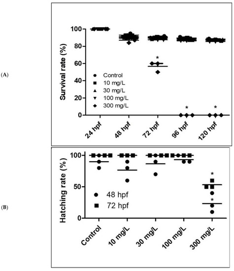
Acrylamide exposure negatively affects survival (A), hatching (B), and heart rate (C). n = 30 for each dose, respectively. * p < 0.01. Error bars represent the standard error of the mean. PHENOTYPE:
|
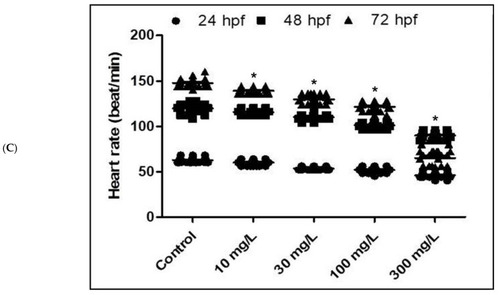
Acrylamide exposure negatively affects survival (A), hatching (B), and heart rate (C). n = 30 for each dose, respectively. * p < 0.01. Error bars represent the standard error of the mean. |
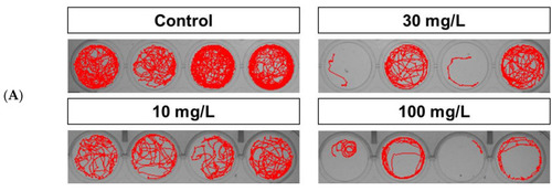
Behavioral effects following a 5-day exposure to acrylamide. (A). Acrylamide-treated wild-type zebrafish larvae exhibited impaired locomotor activity (circular movement and decreased swimming speed and distance traveled) that was concentration-dependent. (B). Distance traveled. (C). Swimming speed. * p < 0.01. Error bars represent the standard error of the mean. |
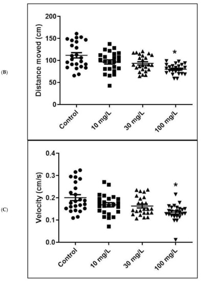
Behavioral effects following a 5-day exposure to acrylamide. (A). Acrylamide-treated wild-type zebrafish larvae exhibited impaired locomotor activity (circular movement and decreased swimming speed and distance traveled) that was concentration-dependent. (B). Distance traveled. (C). Swimming speed. * p < 0.01. Error bars represent the standard error of the mean. |
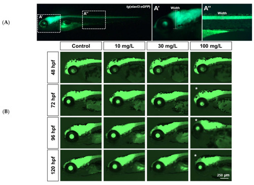
Acrylamide-induced neurotoxicity in transgenic |
Acrylamide-induced neurotoxicity in transgenic |
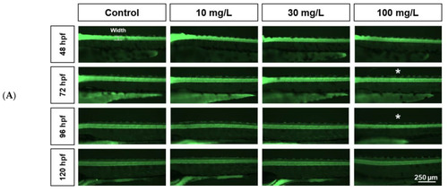
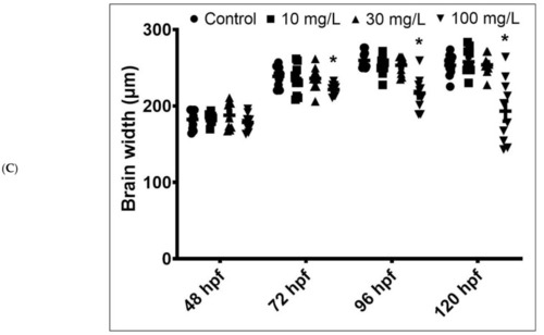
Comparison of spinal cord neuronal width between controls and acrylamide-treated zebrafish. ( PHENOTYPE:
|
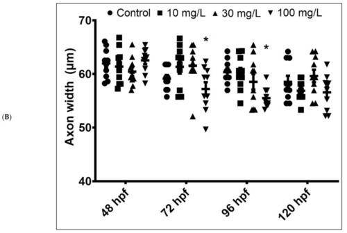
Comparison of spinal cord neuronal width between controls and acrylamide-treated zebrafish. ( |