
- Title
-
Study on Anti-Inflammatory Effect and Major Anti-Inflammatory Components of PSORI-CM02 by Zebrafish Model
- Authors
- Yuan, X., Huang, L., Lei, J., Long, Y., Li, C.
- Source
- Full text @ Evid. Based Complement. Alternat. Med.
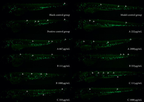
Phenotype map of the effects of each experimental group on zebrafish inflammation. Fluorescence microscopy was used to reveal neutrophils in PHENOTYPE:
|

Phenotype of ink phagocytosis by macrophages in the zebrafish brain. Statistical area is in the black dotted line frame. The white arrow indicates the macrophage that has swallowed ink. Because it has swallowed ink, the color of macrophage is darker than that of normal macrophage. |