Image
Figure Caption
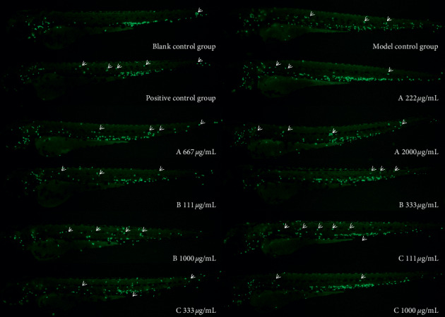
Figure 1
Phenotype map of the effects of each experimental group on zebrafish inflammation. Fluorescence microscopy was used to reveal neutrophils in
Figure Data
Acknowledgments
This image is the copyrighted work of the attributed author or publisher, and
ZFIN has permission only to display this image to its users.
Additional permissions should be obtained from the applicable author or publisher of the image.
Full text @ Evid. Based Complement. Alternat. Med.

