FIGURE SUMMARY
Title

Fundc1 is necessary for proper body axis formation during embryogenesis in zebrafish

Authors
Xu, G., Shen, H., Nibona, E., Wu, K., Ke, X., Al Hafiz, M.A., Liang, X., Zhong, X., Zhou, Q., Qi, C., Zhao, H.
Source
Full text @ Sci. Rep.

ZFIN is incorporating published figure images and captions as part of an ongoing project. Figures from some publications have not yet been curated, or are not available for display because of copyright restrictions.

EXPRESSION / LABELING:
Gene:
Fish:
Anatomical Terms:
Stage Range: 1-cell to Adult

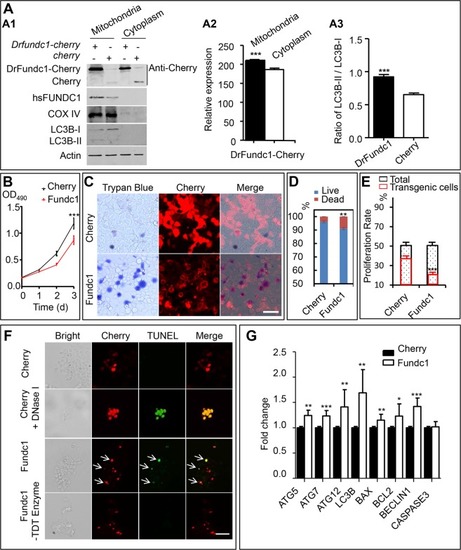
DrFundc1 reduced cell viability while inducing autophagy and apoptosis in transgenic 293 T cells. (
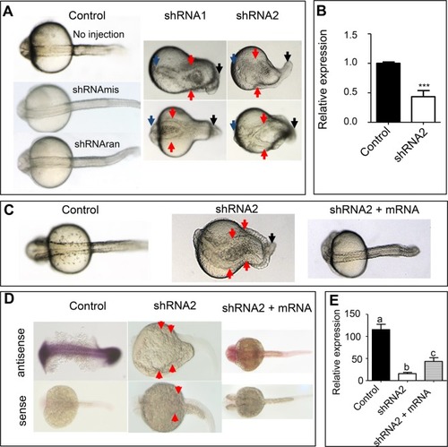
A) Western blotting of proteins extracted from mitochondria and cytoplasm from transgenic 293 T cells transfected with pCS2 + -Drfundc1-Cherry-His as well as pCS2 + -Cherry plasmids. Antibodies used were anti-Cherry (detecting Drfundc1-Cherry-His and Cherry), anti-FUNDC1, anti-COX IV, anti-LC3B, and anti-β-ACTIN. (A1) Results of Western blotting. (A2) Gray-scale analyses of DrFundc1 using ImageJ in mitochondria and cytoplasm of cells transfected with pCS2 + -Drfundc1-Cherry-His. (A3) LC3B-II:LC3B-I ratio in mitochondria of transgenic cells transfected with pCS2 + -Drfundc1-Cherry-His and pCS2 + -Cherry. (B) DrFundc1 decreased viability of 293 T cells, measured by MTT assay. (C-D) DrFundc1 increased 293 T cell mortality, measured by Trypan Blue staining. (E) DrFundc1 decreased cell proliferation in transgenic cells, detected using BrdU incorporation. (F) DrFundc1 led to apoptosis of 293 T cells, detected using TUNEL assay. Arrows show apoptotic cells. Red indicates DrFundc1-Cherry or Cherry, while green indicates TUNEL-positive cells. Positive control: Cherry + DNase I. Negative control: incubation without TdT enzyme. (G) Expressional fold change of autophagy- and apoptosis-related genes, detected using qRT-PCR. β-ACTIN was used as an internal control. Significant differences between cells transfected with different plasmids are shown as asterisks. *P < 0.05; **P < 0.01; ***P < 0.001.

Knockdown of Drfundc1 caused abnormal body axis development in zebrafish embryos (A,B), which was repaired by Drfundc1 mRNA (CE). (A) Midline bifurcation and headless embryos caused by microinjection of Drfudnc1 shRNA1 or shRNA2 into zygotes at 24–28 hpf. (B) shRNA2 significantly decreased endogenous Drfundc1 in the embryos (***P < 0.001), as measured by qRT-PCR with normalization by β-actin. (C) Co-injection of Drfundc1 mRNA repaired defects caused by shRNA2. Before injection, 200 pg/nL of shRNA2 was mixed with 400 pg/nL of mRNA. Control embryos were not injected. (D) WISH results of Drfundc1 in embryos with different treatments. (E) Gray-scale analysis of WISH results of Drfundc1 (D), measured using ImageJ. Letters a, b, and c indicate significance (P < 0.05). All embryos were oriented with a dorsal view. Arrows point to abnormal sites in the embryos.

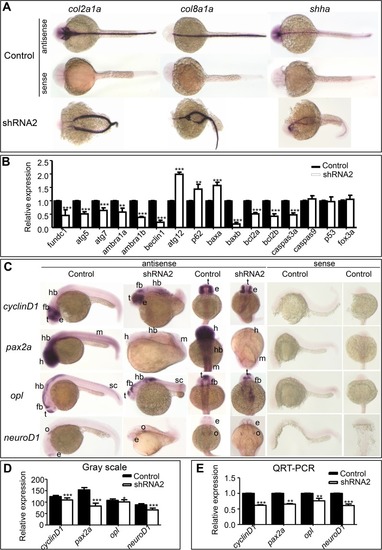
Expression of genes related to body axis formation, autophagy, and apoptosis. (A) WISH of col2a1a, col8a1a, and shha in embryos at 24–28 hpf. (B) Expression of autophagy- and apoptosis-related genes in embryos at 24–28 hpf, detected using qRT-PCR. β-actin was used as an internal control. (C) WISH of cyclinD1, pax2a, opl, and neuroD1, which are involved in neural system development. (D) Gray-scale analysis of WISH results of genes in (C), whose signal strength was measured using ImageJ. (E) Expression of cyclinD1, pax2a, opl, and neuroD1 in embryos, detected using qRT-PCR, was normalized using β-actin.

Acknowledgments
This image is the copyrighted work of the attributed author or publisher, and ZFIN has permission only to display this image to its users. Additional permissions should be obtained from the applicable author or publisher of the image. Full text @ Sci. Rep.