Sequences and predicted structures of zebrafish metalloproteinase 11 (Mmp11) and tissue inhibitors of metalloproteinase-4 (Timp4) paralogues exhibit both conserved and divergent features. (A) Inferred amino acid sequences of zebrafish Mmp11a and Mmp11b paralogues aligned with human MMP11 for reference. Secretory signals are boxed in light blue, propeptide in purple (with cysteine switch motif highlighted in bold and furin recognition sequence underlined), catalytic domain in red (with S-loop in orange, zinc binding motif in bold italic, and specificity loop in light blue), and the carboxyl hemopexin domain is boxed in green. (B) Structural homology models of zebrafish Mmp11a and Mmp11b rendered as ribbon diagrams and colored as in the sequence alignment. (C) Inferred amino acid sequences of zebrafish Timp4a and Timp4b paralogues aligned with human TIMP4 for reference. Secretory signals are boxed in light blue, the N-terminal domain in blue (with the seven-residue charged insertion of Timp4a highlighted in orange), and the C-terminal domain in red. (D) Structural homology models of zebrafish Timp4a and Timp4b rendered as ribbon diagrams and colored as in the sequence alignment.

Schematic illustration of the construction of EMMA–Mmp11a. The coding sequence of zebrafish mmp11a 3′ to the secretory signal was amplified using primers with Gateway recombination sites, recombined into a donor vector, and from there into the EMMA destination vector. This allows the expression of a doubly epitope-tagged protein, whereby loss of co-localization of the HA and GFP epitopes signifies proteolytic activation of the protease by endogenous mechanisms [25].

Mmp11a is activated by furin except in the nucleus. (A) Myocytes expressing EMMA–Mmp11a remove the hemagglutinin (HA)-tagged propeptide (red channel), leaving only the carboxyl terminal GFP tag (green channel) as the construct traverses the secretory pathway, and also in the nucleus. This is more clearly visible in the false color heat map showing the strength of the GFP signal relative to the HA signal (A′). (B) Epithelial cells expressing EMMA–Mmp11a also remove the propeptide as the construct is secreted and released extracellularly, as well as in the nucleus. (B′) Heat maps of the relative abundance of GFP vs. HA clearly illustrate the activation of the EMMA–Mmp11a construct as it is secreted, as well as within the nuclei. (C) Myocytes expressing EMMA–Mmp11a in the presence of furin inhibitor only remove the propeptide in the nuclei. (C′) Heat maps of myocytes expressing the construct in the presence of the furin inhibitor show relatively unimpaired proteolytic removal of the propeptide in the nuclei, but dramatically reduced activation in the secretory pathway and extracellularly. (D) Epithelial cells expressing the construct in the presence of furin inhibitor also show dramatically reduced activation. (D′) Heat maps of epithelial cells in the presence of furin inhibitor, showing negligable activation of the contstruct. (E) Immunoblots of embryo homogenates expressing either GFP alone (flk:GFP), or EMMA–Mmp11a, in the presence or absence of furin inhibitor, probed with anti-GFP. Activation of the construct is clearly detectable in embryos 6 h post heat shock (hpHS) in the absence of inhibitor. In the presence of inhibitor, some activation is detectable 3 hpHS, but by 6 hpHS activation, appears completely abolished. Silver staining of a replicate gel shows comparable protein loads. Scale bars are 10 µm.

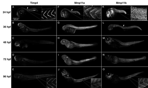
Timp4 and Mmp11 paralogues accumulate dynamically during development and co-localize at the myotendinous junctions (MTJs). Composite confocal projections of 24, 36, 48, 72 and 96 hpf embryos labeled with antibodies against Timp4, Mmp11a, or Mmp11b. (AE) Timp4 immunoreactivity is abundant at the MTJ at all stages examined (arrowhead), and accumulates dynamically in ectodermal epithelia of the head, fin folds, otic vesicle, the posterior notochord and muscle attachments in the jaws and pectoral fins in later stages. (FJ) Mmp11a is present at the MTJs in 24 hpf embryos (F) (arrowhead) but becomes localized within muscle cells in later stages (G,H). (KO) Mmp11b becomes concentrated in MTJs at later stages (LO) (arrowhead). Insets show higher magnification views of MTJs dorsal to the yolk extension in 24 and 96 hpf embryos for comparison. Scale bar = 500 µm.

Timp4 and both Mmp11 paralogues are present in the MTJ at 28 hpf. High-resolution confocal micrographs of MTJs in the trunk skeletal musculature dorsal to the yolk extension of 28 hpf embryos stained with antibodies against (A) Mmp11a, (B) Mmp11b, (C) Timp4, and (D) Laminin (green) as well as anti-α-actinin (red) reveal that both Mmp11 paralogues are detectable in MTJs at this stage, and that they accumulate in the periphery of the MTJ (arrowheads), whereas Timp4 accumulates in the core of the MTJ. Scale bars are 10 µm. Anterior to the left in all images.

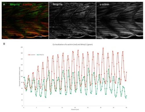
Mmp11a localizes to the Z-discs of skeletal muscle sarcomeres. (A) Confocal micrographs through the trunk musculature of a 48 hpf embryo double labeled with anti-Mmp11a (green) and anti-α-actinin (red) reveal co-localization of the antigens. (B) Quantification of signal intensity in both channels reveals precise co-localization of Mmp11a with α-actinin. Scale bar 50 µm. Anterior to the left.

EXPRESSION / LABELING:
Gene:
Antibodies:
Fish:
Anatomical Term:
Stage: Long-pec

Schematic representation of hypothesized activities and interactions between Mmp11 and Timp4 paralogues at early and late MTJs. The hemopexin-like domain of Mmp11a interacts with the C-terminal domain of Timp4b in the early MTJ, facilitating the degradation of fibronectin. In the mature MTJ, Mmp11b is inhibited by Timp4a, constraining its activity in the maintenance of the ECM, while Mmp11a localizes to the Z-discs of the sarcomeres within the myocytes.

Acknowledgments
This image is the copyrighted work of the attributed author or publisher, and ZFIN has permission only to display this image to its users. Additional permissions should be obtained from the applicable author or publisher of the image. Full text @ J Dev Biol