
- Title
-
2,4-Dinitrotoluene (DNT) Perturbs Yolk Absorption, Liver Development and Lipid Metabolism/Oxygen Transport Gene Expression in Zebrafish Embryos and Larvae
- Authors
- Xiong, J., Sha, H., Zhou, H., Peng, L., Wu, L., Qiu, Y., Wang, R., Hu, X.
- Source
- Full text @ Int. J. Mol. Sci.
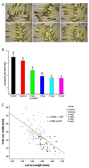
Morphogenesis of zebrafish larvae exposed to different concentrations of 2,4-Dinitrotoluene (2,4-DNT). A: The morphology of larvae exposed to 2,4-DNT from 2 hpf (hours post-fertilzation) to 3 dpf (days post-fertilization). The pictures were taken with the stereomicroscope in 3× magnification. The larvae show light skin, yolk sac accumulation and pericardial edema. The yellow and blue arrows indicate the yolk sac and pericardial cavity, respectively. B: Larvae/yolk sac areas in the control and 2,4-DNT-treated larvae. Bars that do not share a common lower-case letter are significantly different between the treatments (p < 0.05). C: Relationship between the width of the yolk sac and the total length of the larvae. |
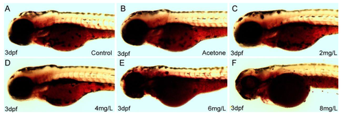
Oil red O (ORO) staining of representative larvae is shown in the left lateral view following different treatments at 3 dpf. A: Control larvae at 3 dpf; B: Vehicle control larvae treated with 0.05 mL/L acetone at 3 dpf; C: 2,4-DNT 2 mg/L-treated larvae at 3 dpf; D: 2,4-DNT 4 mg/L-treated larvae at 3 dpf; E: 2,4-DNT 6 mg/L-treated larvae at 3 dpf; F: 2,4-DNT 8 mg/L-treated larvae at 3 dpf. The pictures were taken with the stereomicroscope in 5× magnification |
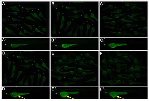
Yolk malabsorption in Tg(apop:GFP) larvae exposed to 2,4-DNT at 3 dpf. A: blank control; B: vehicle control (0.05 mL/L acetone); C: 2 mg/L 2,4-DNT; D: 4 mg/L 2,4-DNT; E: 6 mg/L 2,4-DNT; F: 8 mg/L 2,4-DNT. The yellow arrows indicate yolk sac accumulation. The pictures for A-F were taken with the stereomicroscope in 2.5× magnification, and A’-F’ were in 3.5× magnification. |
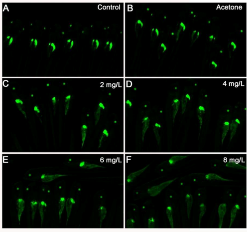
2,4-DNT inhibited the development of hepatic primordium in Tg(apop:GFP) larvae at 5 dpf. A: blank control; B: vehicle control (0.05 mL/L acetone); C: 2 mg/L 2,4-DNT; D: 4 mg/L 2,4-DNT; E: 6 mg/L 2,4-DNT; F: 8 mg/L 2,4-DNT; G–L: left view; G’–L’: right view; G’’–L’’: dorsal view. The pictures for A-F were taken with the stereomicroscope in 3.5× magnification, and G-L, G’–L’ and G’’–L’ were in 4.5× magnification. |
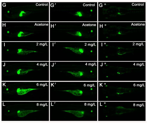
2,4-DNT inhibited the development of hepatic primordium in Tg(apop:GFP) larvae at 5 dpf. A: blank control; B: vehicle control (0.05 mL/L acetone); C: 2 mg/L 2,4-DNT; D: 4 mg/L 2,4-DNT; E: 6 mg/L 2,4-DNT; F: 8 mg/L 2,4-DNT; G–L: left view; G’–L’: right view; G’’–L’’: dorsal view. The pictures for A-F were taken with the stereomicroscope in 3.5× magnification, and G-L, G’–L’ and G’’–L’ were in 4.5× magnification.
|