
Fig. 2
- ID
- ZDB-IMAGE-190822-46
- Publication
- Xiong et al., 2019 - 2,4-Dinitrotoluene (DNT) Perturbs Yolk Absorption, Liver Development and Lipid Metabolism/Oxygen Transport Gene Expression in Zebrafish Embryos and Larvae
- All Figures
- Figures for Xiong et al., 2019
|
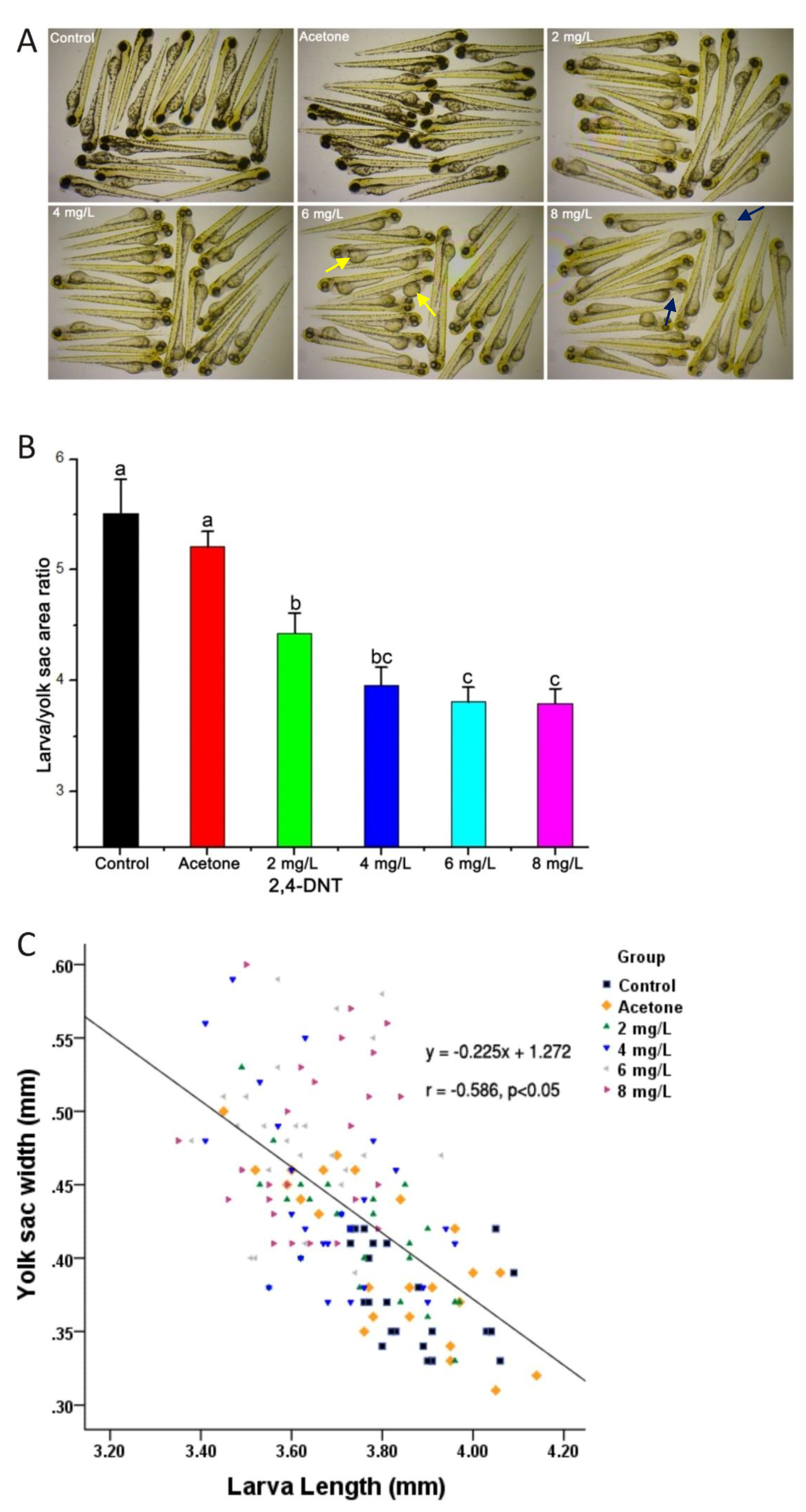
Fig. 2
Morphogenesis of zebrafish larvae exposed to different concentrations of 2,4-Dinitrotoluene (2,4-DNT). A: The morphology of larvae exposed to 2,4-DNT from 2 hpf (hours post-fertilzation) to 3 dpf (days post-fertilization). The pictures were taken with the stereomicroscope in 3× magnification. The larvae show light skin, yolk sac accumulation and pericardial edema. The yellow and blue arrows indicate the yolk sac and pericardial cavity, respectively. B: Larvae/yolk sac areas in the control and 2,4-DNT-treated larvae. Bars that do not share a common lower-case letter are significantly different between the treatments (p < 0.05). C: Relationship between the width of the yolk sac and the total length of the larvae.