
- Title
-
White LED Light Exposure Inhibits the Development and Xanthophore Pigmentation of Zebrafish Embryo
- Authors
- Üstündağ, Ü.V., Çalıskan-Ak, E., Ateş, P.S., Ünal, İ., Eğilmezer, G., Yiğitbaşı, T., Ata Alturfan, A., Emekli-Alturfan, E.
- Source
- Full text @ Sci. Rep.
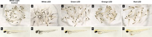
Representative images of the zebrafish embryos at 72 hpf. Examples of individual phenotypes are given in A1–E1. PHENOTYPE:
|

Effects of White LED light pigmentation in zebrafish embryos. (A) Synchronized embryos (n = 20) were exposed to White LED light and White light control. The effects on zebrafish pigmentation were observed under a stereomicroscope at 3 dpf. The pigmentation area density in the treated embryos was normalized to that of the control embryos using the ImageJ software. *p < 0.05, compared to the control. PHENOTYPE:
|

ZFIN is incorporating published figure images and captions as part of an ongoing project. Figures from some publications have not yet been curated, or are not available for display because of copyright restrictions. |
|
ZFIN is incorporating published figure images and captions as part of an ongoing project. Figures from some publications have not yet been curated, or are not available for display because of copyright restrictions. PHENOTYPE:
|
|
ZFIN is incorporating published figure images and captions as part of an ongoing project. Figures from some publications have not yet been curated, or are not available for display because of copyright restrictions. PHENOTYPE:
|
|
ZFIN is incorporating published figure images and captions as part of an ongoing project. Figures from some publications have not yet been curated, or are not available for display because of copyright restrictions. EXPRESSION / LABELING:
PHENOTYPE:
|
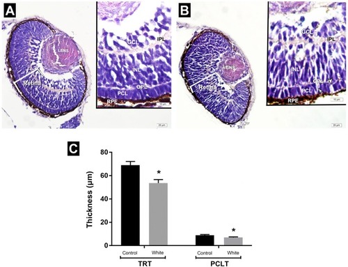
(A,B) Representative photomicrographs of the eyes of zebrafish embryos in the white light control and white LED light exposed groups at 72 hpf. White light control group. (A) Retina with regular layers. White LED light group. (B) disorganization of photoreceptor cells including spaces among cells. (C) Assesment of measurement of the thickness of total retina and photoreceptor cell layer. INL: Inner nuclear layer; GCL: Ganglion cell layer; PCL: Photoreceptor cell layer; IPL: Inner plexiform layer; OPL: Outer plexiform layer; TRT: Total retinal thickness; PCLT: Photoreceptor cell layer thickness; HE staining. PHENOTYPE:
|
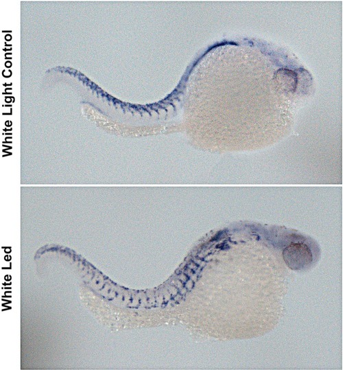
In situ hybridization results revealed decreased crestin expression in white LED exposed group when compared with the white light control group. |

Representative images of the zebrafish embryos at 72 hpf. Examples of individual phenotypes are given in A1–E1. |