
- Title
-
β-Glucan-Producing Pediococcus parvulus 2.6: Test of Probiotic and Immunomodulatory Properties in Zebrafish Models.
- Authors
- Pérez-Ramos, A., Mohedano, M.L., Pardo, M.Á., López, P.
- Source
- Full text @ Front Microbiol
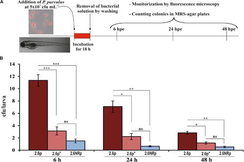
Zebrafish gut colonization by P. parvulus 2.6p and 2.6NRp strains. (A) The experimental procedure is depicted. Four-days-old larvae were colonized during 18 h by 2.6p with (2.6p∗) or without β-glucan removal or 2.6NRp strains. (B) The pediococcal colonies presented inside the larvae were analyzed at 6, 24, and 48 h after the bacteria were removed from the incubation buffer (hours post exposure, hpe). Differences between conditions were evaluated at each point time. Statistical significances are represented by ∗P ≤ 0.05, ∗∗P ≤ 0.01, ∗∗∗P ≤ 0.001, and ns (P > 0.05). |

Fluorescence microscopy images of zebrafish gut colonization by P. parvulus 2.6p and 2.6NRp strains. Representative images taken at 6, 24, and 48 hpe are depicted. White arrows mark the red fluorescence signal emitted by the bacterial cells inside the zebrafish gut. |

Protective effect of P. parvulus 2.6p and 2.6NRp strains against V. anguillarum NB10[pOT11] zebrafish infection. (A) The experimental procedure. Before infection with V. angillarum, 4-days-old larvae were colonized during 18 h either by 2.6p with (2.6p∗) or without β-glucan removal or by 2.6NRp strains. Also, as a control, a group of larvae was incubated in the absence of bacteria during 18 h prior infection. (B) Cumulative mortality due to Vibrio infection was analyzed at 24, 48, and 72 h after addition of the pathogen (hours post infection, hpi). Differences between conditions were evaluated at each point time. Statistical significances are represented by different letters that mean P ≤ 0.05. |
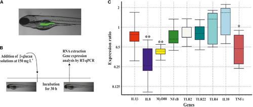
Immunomodulation of gnotobiotic zebrafish larvae by the P. parvulus 2.6 β-glucan. (A) DTAF-labeled β-glucan was detected inside the zebrafish gut. (B) The experimental procedure for the immunomodulation assay. (C) Relative expression values of the genes involved in inflammation. The genes actin-β and elongation factor-1 were used as housekeeping to normalize the values. Statistical significances are represented by ∗P ≤ 0.05 and ∗∗ P ≤ 0.01. |
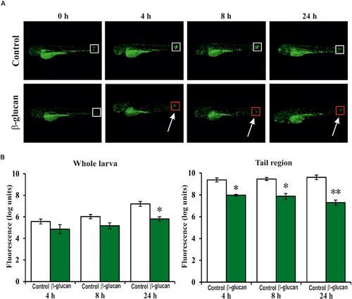
Anti-inflammatory effect of the P. parvulus 2.6 β-glucan in an induced inflammation model of the Tg(mpx:GFP) zebrafish line. The inflammation was induced by cutting off the apical region of the tail. (A) Images of representative larvae treated and untreated with the β-glucan are depicted. Squares mark the areas of inflammation, and the colors indicate regions (red) with less migration of neutrophils than others (white). (B) The green fluorescence emitted by the neutrophils was quantified in the whole larvae and in their tails regions. The fluorescence values detected during the inflammation procedure were expressed as increased percentages of the values detected at 0 h, and represented as logarithms. Statistical significances between groups are represented by ∗P ≤ 0.05 and ∗∗P ≤ 0.01. |