
- Title
-
The progestin norethindrone affects sex differentiation and alters transcriptional profiles of genes along the hypothalamic-pituitary-gonadal and hypothalamic-pituitary-adrenal axes in juvenile zebrafish Dario renio
- Authors
- Hou, L.P., Chen, H., Tian, C.E., Shi, W.J., Liang, Y., Wu, R.R., Fang, X.W., Zhang, C.P., Liang, Y.Q., Xie, L.
- Source
- Full text @ Aquat. Toxicol.
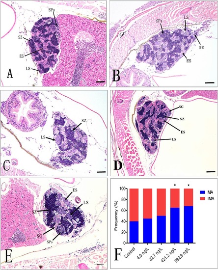
Different germ cell cysts observed during the spermatogenic process in zebrafish. (A): Representative section of a control zebrafish testis with spermatozoa at 65 dpf. (B), (C), (D), and (E): Representative sections of zebrafish testis at 65dpf in the four different NET treatments. (F): Proportion of IMA and MA in the testis. Abbreviations used: ES (early spermatocytes); IMA (immature spermatocytes including spermatogonia and spermatocytes); LS (late spermatocytes); MA (mature spermatocytes including spermatids and spermatozoa); SG (spermatogonia); SP (spermatids); SZ (spermatozoa). Scale bar = 50 μm. Statistically significant differences from the solvent control are marked with asterisks (*p < 0.05). |
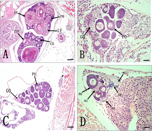
Histological sections of the ovaries of female zebrafish at 65 dpf exposed to different NET concentrations for 45 days (from 20 dpf to 65 dpf). (A) A control ovary showing several developmental stages of follicular cells. (B) An ovary of fish exposed to 4.0 ng/L of NET. (C) An ovary of fish exposed to 32.7 ng/L of NET. (D) An ovary of fish exposed to 421.3 ng/L of NET. (E) An ovary of fish exposed to 892.9 ng/L of NET. (F) The frequency of female with spermatogonia in ovaries. Inter-sex in the zebrafish observed in all NET treatments, showing several spermatogonia and primary spermatozoa in the mature ovary. Abbreviations used: AF (atretic follicle); CO (corticolar alveolar); EV (early vitellogenic oocyte); GO (gonocyte); PO (perinuclear oocyte); SG (spermatogonia); SZ (spermatozoa); LV (late vitellogenic oocyte). Scale bar = 50 μm. |