
|
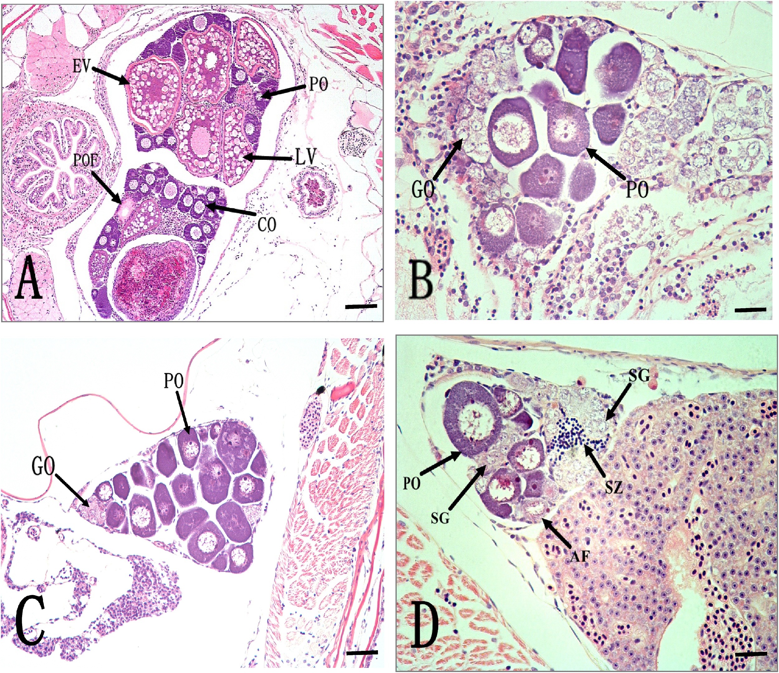
Fig. 3
Histological sections of the ovaries of female zebrafish at 65 dpf exposed to different NET concentrations for 45 days (from 20 dpf to 65 dpf). (A) A control ovary showing several developmental stages of follicular cells. (B) An ovary of fish exposed to 4.0 ng/L of NET. (C) An ovary of fish exposed to 32.7 ng/L of NET. (D) An ovary of fish exposed to 421.3 ng/L of NET. (E) An ovary of fish exposed to 892.9 ng/L of NET. (F) The frequency of female with spermatogonia in ovaries. Inter-sex in the zebrafish observed in all NET treatments, showing several spermatogonia and primary spermatozoa in the mature ovary. Abbreviations used: AF (atretic follicle); CO (corticolar alveolar); EV (early vitellogenic oocyte); GO (gonocyte); PO (perinuclear oocyte); SG (spermatogonia); SZ (spermatozoa); LV (late vitellogenic oocyte). Scale bar = 50 μm.