
- Title
-
The function of tcf3 in medaka embryos: efficient knockdown with pePNAs
- Authors
- Doenz, G., Dorn, S., Aghaallaei, N., Bajoghli, B., Riegel, E., Aigner, M., Bock, H., Werner, B., Lindhorst, T., Czerny, T.
- Source
- Full text @ BMC Biotechnol.

tcf3 expression in medaka embryonic development. Whole mount in situ hybridization experiments were performed in wild type embryos at the indicated stages, using a digoxigenin-labelled RNA probe against tcf3. Embryos are shown in dorsal view, anterior at the top. (f-h) Flat mounts; (f'-h') tail view. During gastrulation (a-c) tcf3 is expressed throughout the epiblast and the embryonic body (c-d). In neurula the expression becomes restricted to the head (d,d') from where it spreads caudally and divides into a forebrain, midbrain, and hindbrain section (e;e'). A gap in expression at the mid-hindbrain boundary (f-h, arrowhead) is closed at later stages (h). In later stages, expression is observed throughout the entire body (g-h'). Scale bars 100 μm; a, a-c; f, f-f′; g, g-h′. Abbreviations: st, stage |

Loss of function phenotypes for the tcf3 gene in medaka. Embryos at the 1-cell stage were co-injected with 300 μM morpholino oligonucleotide (Tcf3MO) and 1 μg/mL FITC-dextran. The pairs b and f, c and g, d and h each show the same embryo at different stages (stage 22, a-d; stage 30, e-h). Control embryos (a,e) were co-injected with 1× Yamamoto’s and FITC-dextran. Pictures of embryos with a fluorescent signal were taken at the indicated stages. Classification was performed after the onset of eye pigmentation, according to the size of the eye: (b,f) weak, slightly reduced size; (c,g) moderate, severe size reduction; (d,h) strong, eye-less. Embryos at stage 16 (i-k, n-o) are shown in dorsal view, anterior to the top; except for (l,p) which are at stage 17 and shown in lateral view. For genotypic analysis (i-p), whole mount in situ hybridization experiments were performed on MO injected embryos and un-injected wild type controls at stage 16 using digoxigenin-labelled RNA probes against pax6 (i,m), pax2 (j,n), gbx1 (k,o), and wnt1 (l,p) at stage 17. (i-k, m-o) Broken lines indicate the outlines of the prospective neural domain estimated from the merged expression patterns formed by pax6, pax2 and gbx1 in wild type embryos (i-k). An anterior shift of the expression domains was observed for all four genes. (l,p; arrowhead) wnt1 expression in the mid-hindbrain boundary. Scale bars 100 μm; a, a-h; i, i-k and m-o; l, l and p. Abbreviations: MO, morpholino oligonucleotide |

tcf3 gain of function phenotypes. a Schematic representation of full length Tcf3, Tcf3 lacking the CtBP binding site (Tcf3[1-434]), and a C-terminally truncated Tcf3 lacking the Groucho/Tle interaction domain (Tcf3[1-434]ΔGro). b-f Embryos at the 1-cell stage were injected with either heat-inducible truncated Tcf3 (Gfp:HSE:Tcf3(1-434); 40 ng/μl) or Tcf3 lacking the Groucho/Tle interaction domain (Gfp:HSE:Tcf3[1-434]ΔGro; 40 ng/μl). Heat induction was performed at stage 14-15. Gfp positive embryos were selected at stage 22 for subsequent whole mount in situ hybridization experiments using digoxigenin-labelled RNA probes against rx2 (c-f). b-e Flat mounts are shown in dorsal view, anterior at the top. Phenotypes were categorized according to their eye size and the expression intensity compared to the control: (c) weak, small eyes, normal expression intensity; (d) small eyes, weak expression; (e), missing eyes and expression. b Heat treated wild type control. f Quantitative results of the injections using rx2 in situ hybridization. Scale bar 100 μm |

Aes-mediated Groucho/Tle loss of function phenotypes. a Schematized presentation of full length Tle protein, the Q-domain, and Aes. For transgenic lines, embryos at the 1-cell stage were injected with 20 ng/μl of DNA (Gfp:HSE:Aes and Gfp:HSE:Q). The F1 generation was heat-induced (10 min, 43.5 °C) at stage 15/16. b-k Embryos are shown in dorsal view, anterior to the top. f-k Flat mounts. c-d Phenotypes of the Aes/Q-mediated Groucho/Tle loss of function were categorized according to their eye phenotype into: (c-c″) weak, smaller eyes tilted towards the midline; (d-d”) moderate, beginning cyclopia anterior; strong (e-e”), cyclopic eye. For whole mount in situ hybridization experiments (f-k), using the indicated digoxigenin-labelled RNA probes, fixation was performed at stage 21; heat treatment occurred as described before. i rx2 expression indicates a reduction in eye size, the eyes are tilted at the anterior end towards the midline (arrowheads); (j) Bf1 expression is reduced; (k) Wnt1 expression indicates a reduction in midbrain size. Instead of a defined expression close to the midline (h, arrows), Aes misexpression resulted in an indistinct expression pattern throughout the midbrain (K, arrows). (b-b″, f-h) control embryos were heat-treated wild type embryos. Scale bars 100 μm; b, b-e”; f, f-k |

Gro/Tle dependence of 1 tcf3 in gain-of-function experiments. Embryos at the 1- cell stage were co-injected with 40 ng/μl of the indicated gfp:HSE:Tcf3 constructs. Heat treatment (10 min, 43.5°C) was applied at stage 14. (A) Statistical overview of the phenotype distribution. Whole mount in situ hybridization experiments for rx2 were performed on embryos at stage 21. (B) dorsal view of a stage 31 embryo with anterior at the top, (C) lateral view with anterior at the left. Arrowheads indicate ectopic otic vesicles, the arrow points to the endogenous otic vesicle. Scale bar 100 μm; B, B and C. |
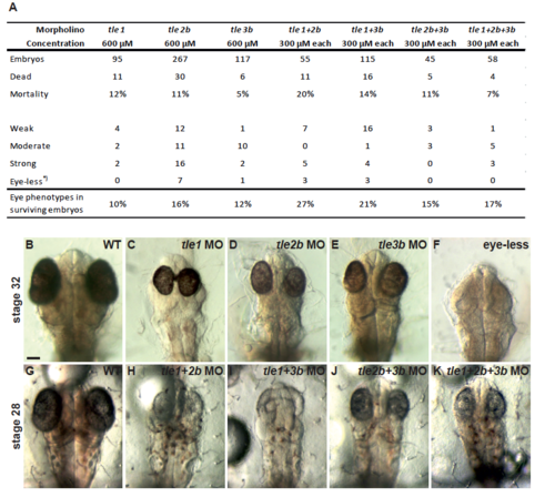
MO-induced gro/tle loss of function phenotypes. Embryos at the 1-cell stage 2 were co-injected with morpholino oligonucleotides and 1 μg/ml FITC-dextran. Injections were 3 performed using either a single morpholino directed against tle1 (C), tle2b (D,F), and tle3b (E), or 4 combinations directed against tle1+2b (H), tle1+3b (I), tle2b+3b (J), and tle1+2b+3b (K). Single 5 morpholino oligonucleotides were injected at a concentration of 600 μM (C-F) and combinatorial 6 injections (H-K) were performed using 300 μM of each MO. Phenotypes of FITC-dextran positive 7 embryos were observed after the beginning of eye pigmentation at stage 32 (B-F) and stage 28 (G-K). 8 (B,G) Wild type control embryos were injected with 1x Yamamoto’s and FITC-dextran. All embryos are 9 shown in dorsal view with anterior at the top. Compared to the wild type controls (B,G), morpholino 10 injected embryos developed smaller eyes that were shifted towards the midline (D,E,J,K) or cyclopic 11 eyes (C,H,I). In rare cases the eyes were lost entirely (E). (A) Phenotypes of FITC-positive embryos 12 were categorized at stage 28-32 into weak and strong phenotypes. Weak phenotypes developed 13 smaller eyes that were shifted towards the midline, whereas strong phenotypes showed cyclopic eyes. 14 Eye-less phenotypes were included into the group of strong phenotypes. Abbreviations: MO, 15 morpholino oligonucleotide; WT, wild type. Scale bar 100 μM. |