
- Title
-
Inhibition of Mitochondrial Division Attenuates Cisplatin-Induced Toxicity in the Neuromast Hair Cells
- Authors
- Vargo, J.W., Walker, S.N., Gopal, S.R., Deshmukh, A.R., McDermott, B.M., Alagramam, K.N., Stepanyan, R.
- Source
- Full text @ Front. Cell. Neurosci.
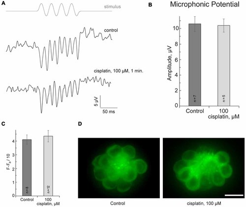
Cisplatin does not affect MET in neuromast hair cells. (A) Neuromast microphonic potentials are not affected after 100-μM-cisplatin application. The top trace shows pressure applied to the stimulating puff pipette. (B) Summary of microphonic potential peak-to-peak amplitudes at twice the stimulus frequency obtained from lateral line neuromasts (controls and after 100-μM-cisplatin application). (C) Summary of fluorescent signal of FM1-43FX in live lateral line neuromasts of control and after 100-μM-cisplatin application. (D) Representative maximum-intensity projection images of FM1-43FX-treated live neuromasts: control and after 100-μM-cisplatin. Data are mean, error bars indicate SEM. n = 5–12 larvae (noted on bar graphs, from three to six clutches) per data point. Scale bar: 10 μm. |

Mdivi-1 protects against cisplatin-induced hair cell death. (A) Concentrations of mitochondrial division inhibitor mdivi-1 between 1–5 μM are well tolerated by zebrafish; whereas 10 μM of mdivi-1 is toxic to hair cells (n = 5 larvae per data point, from three clutches). (B) Application of 3 or 7 μM of mdivi-1 allowed significantly more hair cells to survive treatment with 50 and 100 μM of cisplatin. (C) Representative maximum-intensity projection images of pvalb3b::GFP neuromast hair cells treated with 50-μM-cisplatin and/or 50-μM-mdivi-1 (middle and right images). Data are mean, error bars indicate SEM. **p < 0.001 and *p < 0.05, in comparison to no mdivi-1 treatment within the same cisplatin concentration (see Supplementary Table S1). Scale bar: 10 μm. |