
|
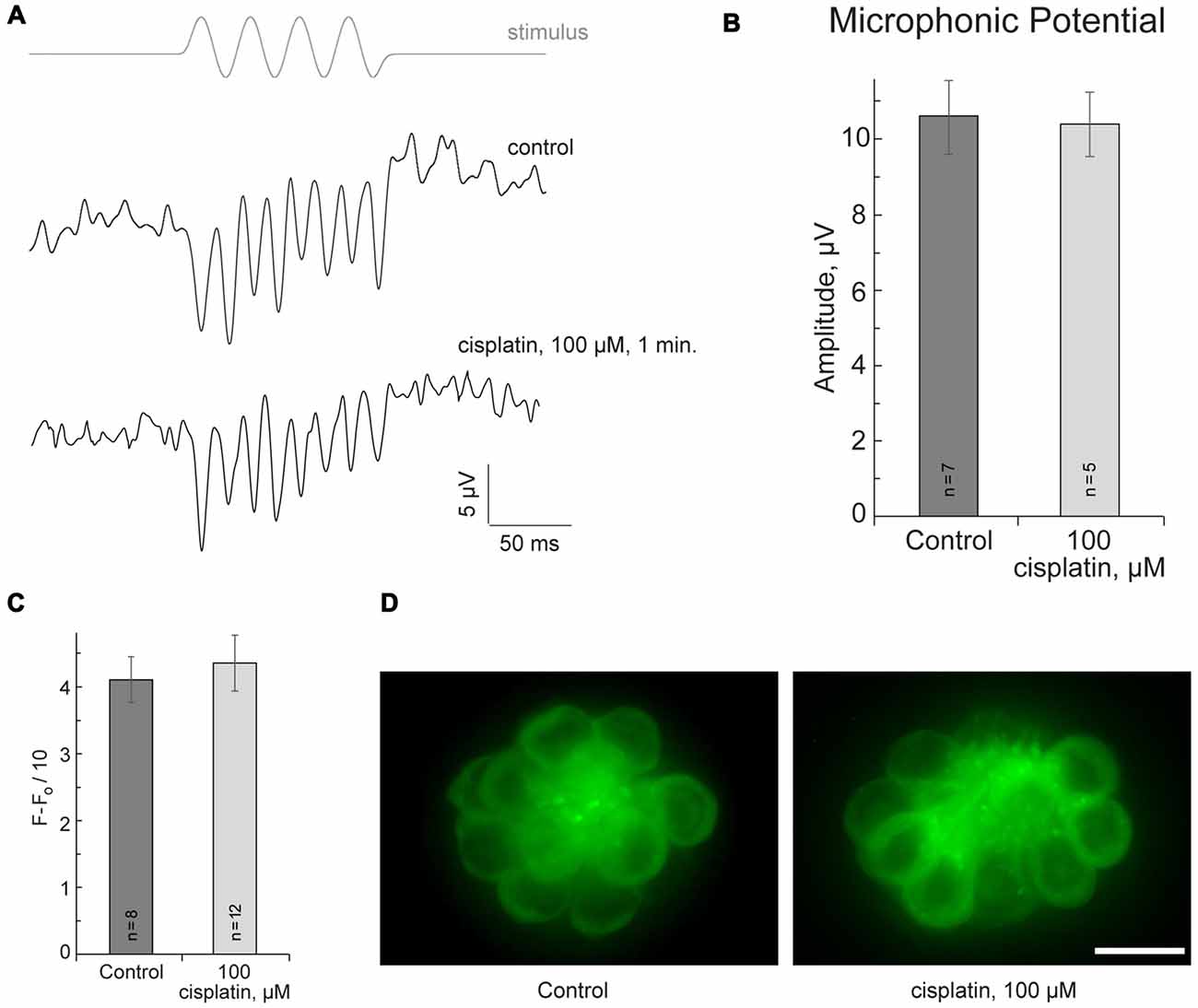
Fig. 2
Cisplatin does not affect MET in neuromast hair cells. (A) Neuromast microphonic potentials are not affected after 100-μM-cisplatin application. The top trace shows pressure applied to the stimulating puff pipette. (B) Summary of microphonic potential peak-to-peak amplitudes at twice the stimulus frequency obtained from lateral line neuromasts (controls and after 100-μM-cisplatin application). (C) Summary of fluorescent signal of FM1-43FX in live lateral line neuromasts of control and after 100-μM-cisplatin application. (D) Representative maximum-intensity projection images of FM1-43FX-treated live neuromasts: control and after 100-μM-cisplatin. Data are mean, error bars indicate SEM. n = 5–12 larvae (noted on bar graphs, from three to six clutches) per data point. Scale bar: 10 μm.