
- Title
-
Francisella noatunensis subspecies noatunensis clpB deletion mutant impairs development of francisellosis in a zebrafish model
- Authors
- Lampe, E.O., Zingmark, C., Tandberg, J.I., Thrane, I.M.P., Brudal, E., Sjöstedt, A., Winther-Larsen, H.C.
- Source
- Full text @ Vaccine

Intravascular injection experiment of zebrafish embryos with green fluorescent F.n.n. wt, F.n.n. ΔclpB and PBS suggests growth attenuation of the deletion mutant. (A) Injections of 102 –103 CFU were administered into the Duct of Cuvier (arrow) of zebrafish embryos (AB wt) and micrographs were made from the tail region (rectangle). (B) Micrographs from the tail region of zebrafish embryos 7 days post injection with green fluorescent F.n.n. gfp (wt), F.n.n. ΔclpB gfp (ΔclpB) and PBS. Only few weakly green fluorescent foci were visible in mutant infected embryos (arrowhead), 10x magnification. (C) Kaplan − Meier curve of cumulative survival of zebrafish embryos injected with F.n.n. gfp, F.n.n. ΔclpB gfp or PBS (control). The difference in cumulative survival between the mutant-injected and the wt-injected embryos was statistically significant (p = .0087). (D) Quantification of F.n.n. genome equivalents by qPCR on gDNA zebrafish embryos injected with F.n.n. gfp or F.n.n. ΔclpB gfp at 0 or 7 days post injection (dpi). Results are presented as mean ± SEM. Bacterial load was significantly higher in wt injected embryos 7 dpi (wt day 0 versus day 7, p = .0089 and ΔclpB day 0 versus day 7, p = .0175 and wt versus ΔclpB day 7, p = .0063). (For interpretation of the references to colour in this figure legend, the reader is referred to the web version of this article.) |
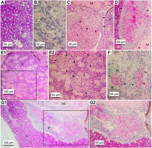
Histology sections of F.n.n. ΔclpB-immunized adult zebrafish before and after challenge with a lethal dose of F.n.n. wt. Sections are stained with Hematoxylin and eosin (HE) or Periodic acid Schiff’s reagent (PAS). PAS stained liver (A) and kidney (B) tissue of zebrafish 27 days after immunization with F.n.n. ΔclpB (one day before challenge). (C-G2) Histology sections from mutant-immunized zebrafish 28 days post challenge (dpc) with F.n.n. wt. (C) A granulomatous process delineated by arrowheads is located in the liver, L next to kidney, K and muscle tissue, M. H&E staining. (D) A large liver granuloma adjacent to muscle tissue, PAS staining. (E1) Overview of HE stained kidney tissue with rectangle showing area magnified in (E2), presenting three localized granulomatous processes (arrowheads). (F) PAS stained section of kidney revealing two granulomas. (G1) Overview of PAS stained tissue with a large granulomatous process localized within pancreatic tissue, P, next to the intestines, Int, with rectangle showing magnified area in (G2). |
Reprinted from Vaccine, 35(52), Lampe, E.O., Zingmark, C., Tandberg, J.I., Thrane, I.M.P., Brudal, E., Sjöstedt, A., Winther-Larsen, H.C., Francisella noatunensis subspecies noatunensis clpB deletion mutant impairs development of francisellosis in a zebrafish model, 7264-7272, Copyright (2017) with permission from Elsevier. Full text @ Vaccine