- Title
-
Aeromonas hydrophila utilizes TLR4 topology for synchronous activation of MyD88 and TRIF to orchestrate anti-inflammatory responses in zebrafish.
- Authors
- Srivastava, N., Shelly, A., Kumar, M., Pant, A., Das, B., Majumdar, T., Mazumder, S.
- Source
- Full text @ Cell Death Discov
|
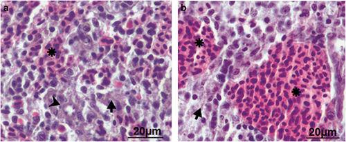
Histological analysis elucidated absence of inflammation in zebrafish spleen. Spleen was removed from control and infected zebrafish and the parenchyma of both was compared (×40). (a) Control zebrafish spleen comprises hematogenous red pulp (asterisk), lymphoid white pulp (arrow) and ellipsoid (arrow head). (b) A. hydrophila-infected fish spleen revealed significant expansion of the red pulp area. |
|
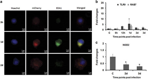
A. hydrophila trafficking in zebrafish. (a) Zebrafish were infected with mCherry-tagged A. hydrophila. Bacterial localization was revealed by immunofluorescence staining of zSPM using Hoechst 33342 and EEA1 at indicated time points (×100). (b, c) Expression of TLR9, RAB7 and NOD2 was determined from spleen of control and infected fishes. Each bar represent the mean of three independent experiments (n=5/experiment) and the error bars represent the standard deviations. C, control; d, days; h, hours; zSPM, zebrafish splenic macrophages; and asterisks '∗' on bars indicate significant difference from control (P<0.05). |
|
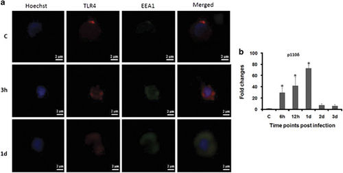
Subcellular distribution of TLR4. (a) zSPM were isolated from control and infected fish, stained with Hoechst 33342, TLR4 and EEA1 antibody for TLR4 internalization study (×100). (b) Expression of p110δ was determined from spleen of control and infected fishes. Each bar represent the mean of three independent experiments (n=5/experiment) and the error bars represent the standard deviations. C, control; d, days; h, hours; zSPM, zebrafish splenic macrophages and asterisks '∗' on bars indicate significant difference from control (P<0.05). |


