Image
Figure Caption
Fig. 6
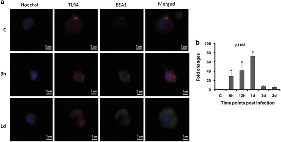
Subcellular distribution of TLR4. (a) zSPM were isolated from control and infected fish, stained with Hoechst 33342, TLR4 and EEA1 antibody for TLR4 internalization study (×100). (b) Expression of p110δ was determined from spleen of control and infected fishes. Each bar represent the mean of three independent experiments (n=5/experiment) and the error bars represent the standard deviations. C, control; d, days; h, hours; zSPM, zebrafish splenic macrophages and asterisks '∗' on bars indicate significant difference from control (P<0.05).
Acknowledgments
This image is the copyrighted work of the attributed author or publisher, and
ZFIN has permission only to display this image to its users.
Additional permissions should be obtained from the applicable author or publisher of the image.
Full text @ Cell Death Discov

