
- Title
-
Mutations in INPP5K, Encoding a Phosphoinositide 5-Phosphatase, Cause Congenital Muscular Dystrophy with Cataracts and Mild Cognitive Impairment
- Authors
- Wiessner, M., Roos, A., Munn, C.J., Viswanathan, R., Whyte, T., Cox, D., Schoser, B., Sewry, C., Roper, H., Phadke, R., Marini Bettolo, C., Barresi, R., Charlton, R., Bönnemann, C.G., Abath Neto, O., Reed, U.C., Zanoteli, E., Araújo Martins Moreno, C., Ertl-Wagner, B., Stucka, R., De Goede, C., Borges da Silva, T., Hathazi, D., Dell'Aica, M., Zahedi, R.P., Thiele, S., Müller, J., Kingston, H., Müller, S., Curtis, E., Walter, M.C., Strom, T.M., Straub, V., Bushby, K., Muntoni, F., Swan, L.E., Lochmüller, H., Senderek, J.
- Source
- Full text @ Am. J. Hum. Genet.
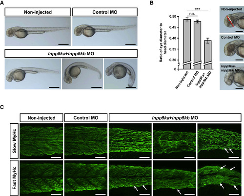
Defects in Zebrafish inpp5ka+inpp5kb Double Morphants at 48 hpf (A) Live embryos injected with control MO, inpp5ka MO, and inpp5kb MO or left untreated (non-injected). More than 95% of living non-injected embryos and embryos injected with control MO showed no macroscopic abnormalities. Images for inpp5ka+inpp5kb morphants represent mild (left, 15% of living embryos), moderate (middle, 24% of living embryos), and severe (right, 44% of living embryos) defects in terms of length and curvature of tails. At least 150 living embryos were counted per condition. Scale bars represent 500 μm. (B) Eye-to-head ratio of zebrafish embryos at 48 hpf. Eye diameter (white line) and head diameter (red line) were measured in the dorsal-ventral axis and ratios were calculated. Scale bars represent 250 μm. Graphs represent mean values of ratios obtained with ten embryos and error bars represent standard deviations. The statistical difference between non-injected and control MO-injected embryos and inpp5ka+inpp5kb double morphants is indicated: n.s. indicates not significant, ∗∗∗p < 0.001 (one-tailed Student’s t test). (C) Whole-mount immunostainings of zebrafish embryos at 48 hpf using antibodies against slow muscle myosin heavy chain (slow MyHc) and fast muscle myosin heavy chain (fast MyHc). Images for inpp5ka+inpp5kb morphants represent mild, moderate, and severe phenotypes (from left to right) as staged macroscopically. inpp5ka+inpp5kb-deficient embryos displayed muscle fiber defects and loss of the chevron shape of somites. The normal straight alignment of slow twitch muscle fibers was disrupted in inpp5ka+inpp5kb double knockdown morphants. Fibers appeared wavy and deformation of myosepta was observed in severe phenotypes (arrows). Staining for fast twitch muscle fibers also showed distortion and defects of fibers and severe alterations of myosepta (arrows). Scale bars represent 50 μm. PHENOTYPE:
|
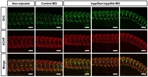
Neuromuscular junction morphology in inpp5ka+inpp5kb morphants. For whole-mount immunofluorescence staining, 48-hpf zebrafish embryos were fixed in 4% paraformaldehyde in PBS at 4°C overnight and blocked for 1 hr at RT in 5% horse serum in PBS, 0.1% Tween-20. Presynaptic motor nerve endings were visualized by incubation with mouse anti-synaptic vesicle protein 2 antibody (SV2, DSHB; 1:200) overnight at 4°C followed by Alexa Fluor 488-conjugated goat anti-mouse IgG antibody for 1 hr at RT. Acetylcholine receptors (AChR) were labelled with Alexa Fluor 594-conjugated α- bungarotoxin (Thermo Fisher Scientific; 1:1,000). Images were captured with a Nikon A1R confocal microscope (Nikon). Images for inpp5ka+inpp5kb morphants represent mild, moderate and severe phenotypes (from left to right) as staged macroscopically. Both in controls and in inpp5ka+inpp5kb morphants, motor axons had made branches into somites where they formed contacts with AChR clusters. Scale bars = 50 μm. PHENOTYPE:
|

ZFIN is incorporating published figure images and captions as part of an ongoing project. Figures from some publications have not yet been curated, or are not available for display because of copyright restrictions. PHENOTYPE:
|