
Fig. S8
- ID
- ZDB-IMAGE-170428-2
- Publication
- Wiessner et al., 2017 - Mutations in INPP5K, Encoding a Phosphoinositide 5-Phosphatase, Cause Congenital Muscular Dystrophy with Cataracts and Mild Cognitive Impairment
- All Figures
- Figures for Wiessner et al., 2017
|
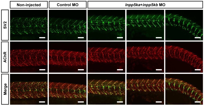
Fig. S8
Neuromuscular junction morphology in inpp5ka+inpp5kb morphants.
For whole-mount immunofluorescence staining, 48-hpf zebrafish embryos were fixed in 4% paraformaldehyde in PBS at 4°C overnight and blocked for 1 hr at RT in 5% horse serum in PBS, 0.1% Tween-20. Presynaptic motor nerve endings were visualized by incubation with mouse anti-synaptic vesicle protein 2 antibody (SV2, DSHB; 1:200) overnight at 4°C followed by Alexa Fluor 488-conjugated goat anti-mouse IgG antibody for 1 hr at RT. Acetylcholine receptors (AChR) were labelled with Alexa Fluor 594-conjugated α- bungarotoxin (Thermo Fisher Scientific; 1:1,000). Images were captured with a Nikon A1R confocal microscope (Nikon). Images for inpp5ka+inpp5kb morphants represent mild, moderate and severe phenotypes (from left to right) as staged macroscopically. Both in controls and in inpp5ka+inpp5kb morphants, motor axons had made branches into somites where they formed contacts with AChR clusters. Scale bars = 50 μm.